HELIX HELIX DETERMINATION METHOD: KABSCH AND SANDER ALGORITHM CHECKED BY HAND ON THE GRAPHICS DISPLAY.
Remark 700
SHEET SHEET SHEET_ID: B-1; DETERMINATION METHOD: KABSCH AND SANDER ALGORITHM CHECKED BY HAND ON THE ...SHEET SHEET SHEET_ID: B-1; DETERMINATION METHOD: KABSCH AND SANDER ALGORITHM CHECKED BY HAND ON THE GRAPHICS DISPLAY.
Mass: 18.015 Da / Num. of mol.: 92 / Source method: isolated from a natural source / Formula: H2O
Compound details
COMPND MOLECULE: PHOTOACTIVE YELLOW PROTEIN. 4-HYDROXYCINNAMYL CHROMOPHORE COVALENTLY LINKED TO ...COMPND MOLECULE: PHOTOACTIVE YELLOW PROTEIN. 4-HYDROXYCINNAMYL CHROMOPHORE COVALENTLY LINKED TO CYSTEINE 69 BY A THIOESTER BOND. TURN KABSCH AND SANDER ALGORITHM CHECKED BY HAND ON THE GRAPHICS DISPLAY.
Has protein modification
Y
Nonpolymer details
4-HYDROXYCINNAMYL CHROMOPHORE, O(C6H4)CH=CH(C=O), IS COVALENTLY LINKED TO CYSTEINE 69 BY A ...4-HYDROXYCINNAMYL CHROMOPHORE, O(C6H4)CH=CH(C=O), IS COVALENTLY LINKED TO CYSTEINE 69 BY A THIOESTER BOND. SPECTROSCOPICALLY, THE CHROMOPHORE APPEARS TO BE NEGATIVELY CHARGED IN THE CONTEXT OF THE FOLDED PROTEIN IN THE DARK STATE. DURING REFINEMENT, CYS 69 AND THE COVALENTLY BOUND CHROMOPHORE WERE TREATED TOGETHER AS ONE RESIDUE.
-
Experimental details
-
Experiment
Experiment
Method: X-RAY DIFFRACTION
-
Sample preparation
Crystal
Density Matthews: 1.9 Å3/Da / Density % sol: 35.17 %
Crystal grow
*PLUS
pH: 7 / Method: vapor diffusion, hanging drop / Details: used as seeds
Components of the solutions
*PLUS
ID
Conc.
Common name
Crystal-ID
Sol-ID
Chemical formula
1
3.2M
1
reservoir
NH4SO4
2
20mM
sodiumphosphate
1
reservoir
3
15mg/ml
protein
1
drop
4
2.5M
1
drop
NH4SO4
5
20mM
sodiumphosphate
1
drop
-
Data collection
Detector
Date: Nov 1, 1991
Radiation
Monochromatic (M) / Laue (L): M / Scattering type: x-ray
In the structure databanks used in Yorodumi, some data are registered as the other names, "COVID-19 virus" and "2019-nCoV". Here are the details of the virus and the list of structure data.
Jan 31, 2019. EMDB accession codes are about to change! (news from PDBe EMDB page)
EMDB accession codes are about to change! (news from PDBe EMDB page)
The allocation of 4 digits for EMDB accession codes will soon come to an end. Whilst these codes will remain in use, new EMDB accession codes will include an additional digit and will expand incrementally as the available range of codes is exhausted. The current 4-digit format prefixed with “EMD-” (i.e. EMD-XXXX) will advance to a 5-digit format (i.e. EMD-XXXXX), and so on. It is currently estimated that the 4-digit codes will be depleted around Spring 2019, at which point the 5-digit format will come into force.
The EM Navigator/Yorodumi systems omit the EMD- prefix.
Related info.:Q: What is EMD? / ID/Accession-code notation in Yorodumi/EM Navigator
Yorodumi is a browser for structure data from EMDB, PDB, SASBDB, etc.
This page is also the successor to EM Navigator detail page, and also detail information page/front-end page for Omokage search.
The word "yorodu" (or yorozu) is an old Japanese word meaning "ten thousand". "mi" (miru) is to see.
Related info.:EMDB / PDB / SASBDB / Comparison of 3 databanks / Yorodumi Search / Aug 31, 2016. New EM Navigator & Yorodumi / Yorodumi Papers / Jmol/JSmol / Function and homology information / Changes in new EM Navigator and Yorodumi
 Movie
Movie Controller


 Open data
 Basic information
 Components
 Keywords
 Function and homology information
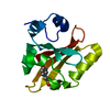
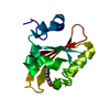
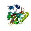
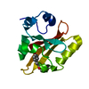
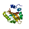
 Halorhodospira halophila (bacteria)
 X-RAY DIFFRACTION / Resolution: 1.4 Å
  Authors
 Citation
 Structure visualization
 Downloads & links
 Other downloads
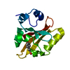


 PDBj
PDBj


 Assembly
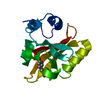

 Mass: 164.158 Da / Num. of mol.: 1 / Source method: obtained synthetically / Formula: C9H8O3
 Mass: 18.015 Da / Num. of mol.: 92 / Source method: isolated from a natural source / Formula: H2O
 Sample preparation
 Processing