 Movie
Movie Controller

[English] 日本語
 Yorodumi
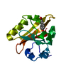
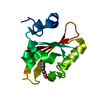
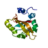
- PDB-2zoh: X-ray Crystal Structure of Photoactive Yellow Protein, Wild type,... -
+ Open data

- Basic information
| Entry | Database: PDB / ID: 2zoh | ||||||
|---|---|---|---|---|---|---|---|
| Title | X-ray Crystal Structure of Photoactive Yellow Protein, Wild type, at 295K | ||||||
 Components Components | Photoactive yellow protein | ||||||
 Keywords Keywords | SIGNALING PROTEIN / PAS / LOV / PHOTORECEPTOR / LIGHT SENSOR / Chromophore / Photoreceptor protein / Receptor / Sensory transduction | ||||||
| Function / homology |  Function and homology information Function and homology informationphotoreceptor activity / phototransduction / regulation of DNA-templated transcription / identical protein binding Similarity search - Function | ||||||
| Biological species |  Halorhodospira halophila (bacteria) Halorhodospira halophila (bacteria) | ||||||
| Method |  X-RAY DIFFRACTION / SYNCHROTRON / MOLECULAR REPLACEMENT / Resolution: 1.25 Å X-RAY DIFFRACTION / SYNCHROTRON / MOLECULAR REPLACEMENT / Resolution: 1.25 Å | ||||||
 Authors Authors | Yamaguchi, S. | ||||||
 Citation Citation | Journal: Proc.Natl.Acad.Sci.USA / Year: 2009 Title: Low-barrier hydrogen bond in photoactive yellow protein Authors: Yamaguchi, S. / Kamikubo, H. / Kurihara, K. / Kuroki, R. / Niimura, N. / Shimizu, N. / Yamazaki, Y. / Kataoka, M. | ||||||
| History |
|
- Structure visualization
| Structure viewer | Molecule: MolmilJmol/JSmol |
|---|
- Downloads & links
-Download
| PDBx/mmCIF format | 2zoh.cif.gz | 70.4 KB | Display | PDBx/mmCIF format |
|---|---|---|---|---|
| PDB format | pdb2zoh.ent.gz | 50.6 KB | Display | PDB format |
| PDBx/mmJSON format | 2zoh.json.gz | Tree view | PDBx/mmJSON format | |
| Others |  Other downloads Other downloads |
-Validation report
| Arichive directory | https://data.pdbj.org/pub/pdb/validation_reports/zo/2zohftp://data.pdbj.org/pub/pdb/validation_reports/zo/2zoh | HTTPS FTP |
|---|
-Related structure data
| Related structure data | 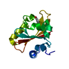 2zoiC  2phyS C: citing same article ( S: Starting model for refinement |
|---|---|
| Similar structure data |


-Links
 PDBj
PDBj


- Assembly
| Deposited unit | 
| ||||||||
|---|---|---|---|---|---|---|---|---|---|
| 1 |
| ||||||||
| Unit cell |
|
-Components
| #1: Protein | Mass: 13888.575 Da / Num. of mol.: 1 Source method: isolated from a genetically manipulated source Source: (gene. exp.) Halorhodospira halophila (bacteria) / Gene: pyp / Plasmid: pET16b / Production host: |
|---|---|
| #2: Chemical | ChemComp-HC4 /   Mass: 164.158 Da / Num. of mol.: 1 / Source method: obtained synthetically / Formula: C9H8O3 Mass: 164.158 Da / Num. of mol.: 1 / Source method: obtained synthetically / Formula: C9H8O3 |
| #3: Water | ChemComp-HOH /  Mass: 18.015 Da / Num. of mol.: 120 / Source method: isolated from a natural source / Formula: H2O Mass: 18.015 Da / Num. of mol.: 120 / Source method: isolated from a natural source / Formula: H2O |
-Experimental details
-Experiment
| Experiment | Method: X-RAY DIFFRACTION / Number of used crystals: 1 |
|---|
- Sample preparation
| Crystal | Density Matthews: 1.9 Å3/Da / Density % sol: 35.24 % |
|---|---|
| Crystal grow | Temperature: 295 K / Method: vapor diffusion, hanging drop, microseeding / pH: 9 Details: AMMONIUM SULPHATE, SODIUM CHLORIDE, SODIUM DIDEUTERIUM PHOSPHATE, DISODIUM DEUTERIUM PHOSPHATE, pH 9.0, VAPOR DIFFUSION, HANGING DROP, MICROSEEDING, temperature 295K |
-Data collection
| Diffraction | Mean temperature: 295 K |
|---|---|
| Diffraction source | Source: SYNCHROTRON / Site: SPring-8  / Beamline: BL41XU / Wavelength: 0.45 / Wavelength: 0.45 Å / Beamline: BL41XU / Wavelength: 0.45 / Wavelength: 0.45 Å |
| Detector | Type: ADSC QUANTUM 315 / Detector: CCD / Date: Dec 3, 2006 |
| Radiation | Monochromator: SI 111 / Protocol: SINGLE WAVELENGTH / Monochromatic (M) / Laue (L): M / Scattering type: x-ray |
| Radiation wavelength | Wavelength: 0.45 Å / Relative weight: 1 |
| Reflection | Resolution: 1.25→57.83 Å / Num. all: 28967 / Num. obs: 28967 / % possible obs: 100 % / Redundancy: 11.1 % / Biso Wilson estimate: 10.11 Å2 / Rmerge(I) obs: 0.058 / Rsym value: 0.058 / Net I/σ(I): 21.9 |
| Reflection shell | Resolution: 1.25→1.32 Å / Redundancy: 11.3 % / Rmerge(I) obs: 0.356 / Mean I/σ(I) obs: 7.3 / Num. unique all: 4176 / Rsym value: 0.356 / % possible all: 100 |
- Processing
| Software |
| |||||||||||||||||||||||||||||||||
|---|---|---|---|---|---|---|---|---|---|---|---|---|---|---|---|---|---|---|---|---|---|---|---|---|---|---|---|---|---|---|---|---|---|---|
| Refinement | Method to determine structure: MOLECULAR REPLACEMENT Starting model: 2PHY Resolution: 1.25→50 Å / Num. parameters: 10353 / Num. restraintsaints: 13011 / Isotropic thermal model: ANISOTROPIC / Cross valid method: FREE R / σ(F): 0 / Stereochemistry target values: Engh & Huber
| |||||||||||||||||||||||||||||||||
| Refine analyze | Num. disordered residues: 10 / Occupancy sum hydrogen: 0 / Occupancy sum non hydrogen: 1107 | |||||||||||||||||||||||||||||||||
| Refinement step | Cycle: LAST / Resolution: 1.25→50 Å
| |||||||||||||||||||||||||||||||||
| Refine LS restraints |
|
