 Movie
Movie Controller

[English] 日本語
 Yorodumi
- PDB-2zoi: Neutron Crystal Structure of Photoactive Yellow Protein, Wild typ... -
+ Open data

- Basic information
| Entry | Database: PDB / ID: 2zoi | ||||||
|---|---|---|---|---|---|---|---|
| Title | Neutron Crystal Structure of Photoactive Yellow Protein, Wild type, at 295K | ||||||
 Components Components | Photoactive yellow protein | ||||||
 Keywords Keywords | SIGNALING PROTEIN / PAS / LOV / PHOTORECEPTOR / LIGHT SENSOR / LBHB / SHB / Chromophore / Photoreceptor protein / Receptor / Sensory transduction | ||||||
| Function / homology |  Function and homology information Function and homology informationphototransduction / photoreceptor activity / regulation of DNA-templated transcription / identical protein binding Similarity search - Function | ||||||
| Biological species |  Halorhodospira halophila (bacteria) Halorhodospira halophila (bacteria) | ||||||
| Method | NEUTRON DIFFRACTION / NUCLEAR REACTOR /  MOLECULAR REPLACEMENT / Resolution: 1.5 Å MOLECULAR REPLACEMENT / Resolution: 1.5 Å | ||||||
 Authors Authors | Yamaguchi, S. | ||||||
 Citation Citation | Journal: Proc.Natl.Acad.Sci.USA / Year: 2009 Title: Low-barrier hydrogen bond in photoactive yellow protein Authors: Yamaguchi, S. / Kamikubo, H. / Kurihara, K. / Kuroki, R. / Niimura, N. / Shimizu, N. / Yamazaki, Y. / Kataoka, M. | ||||||
| History |
|
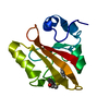
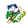
- Structure visualization
| Structure viewer | Molecule: MolmilJmol/JSmol |
|---|
- Downloads & links
-Download
| PDBx/mmCIF format | 2zoi.cif.gz | 63.2 KB | Display | PDBx/mmCIF format |
|---|---|---|---|---|
| PDB format | pdb2zoi.ent.gz | 47 KB | Display | PDB format |
| PDBx/mmJSON format | 2zoi.json.gz | Tree view | PDBx/mmJSON format | |
| Others |  Other downloads Other downloads |
-Validation report
| Arichive directory | https://data.pdbj.org/pub/pdb/validation_reports/zo/2zoiftp://data.pdbj.org/pub/pdb/validation_reports/zo/2zoi | HTTPS FTP |
|---|
-Related structure data
| Related structure data | 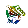 2zohSC S: Starting model for refinement C: citing same article ( |
|---|---|
| Similar structure data |


-Links
 PDBj
PDBj


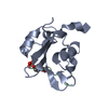
- Assembly
| Deposited unit | 
| ||||||||
|---|---|---|---|---|---|---|---|---|---|
| 1 |
| ||||||||
| Unit cell |
|
-Components
| #1: Protein | Mass: 13888.575 Da / Num. of mol.: 1 Source method: isolated from a genetically manipulated source Source: (gene. exp.) Halorhodospira halophila (bacteria) / Gene: PYP / Plasmid: pET16b / Production host: |
|---|---|
| #2: Chemical | ChemComp-HC4 /   Mass: 164.158 Da / Num. of mol.: 1 / Source method: obtained synthetically / Formula: C9H8O3 Mass: 164.158 Da / Num. of mol.: 1 / Source method: obtained synthetically / Formula: C9H8O3 |
| #3: Chemical | ChemComp-DOD /   Mass: 18.015 Da / Num. of mol.: 73 / Source method: isolated from a natural source / Formula: D2O Mass: 18.015 Da / Num. of mol.: 73 / Source method: isolated from a natural source / Formula: D2O |
-Experimental details
-Experiment
| Experiment | Method: NEUTRON DIFFRACTION / Number of used crystals: 1 |
|---|
- Sample preparation
| Crystal grow | Temperature: 295 K / Method: vapor diffusion, hanging drop, microseeding / pH: 9 Details: AMMONIUM SULPHATE, SODIUM CHLORIDE, SODIUM DIDEUTERIUM PHOSPHATE, DISODIUM DEUTERIUM PHOSPHATE, pH 9.0, VAPOR DIFFUSION, HANGING DROP, MICROSEEDING, temperature 295K |
|---|
-Data collection
| Diffraction | Mean temperature: 295 K |
|---|---|
| Diffraction source | Source: NUCLEAR REACTOR / Wavelength: 2.6 / Wavelength: 2.6 Å |
| Detector | Type: MACSCIENCE DIP SCANNER FOR NEUTRONS / Detector: IMAGE PLATE / Date: Mar 31, 2007 |
| Radiation | Monochromator: ELASTICALLY-BENT PERFECT SI(111) / Protocol: SINGLE WAVELENGTH / Monochromatic (M) / Laue (L): M / Scattering type: x-ray |
| Radiation wavelength | Wavelength: 2.6 Å / Relative weight: 1 |
| Reflection | Resolution: 1.5→100 Å / Num. obs: 15293 / % possible obs: 89.6 % / Redundancy: 2.6 % / Biso Wilson estimate: 9.644 Å2 / Rmerge(I) obs: 0.109 / Net I/σ(I): 7.4 |
| Reflection shell | Resolution: 1.5→1.55 Å / Redundancy: 2 % / Rmerge(I) obs: 0.324 / Mean I/σ(I) obs: 2.91 / Num. unique all: 1234 / % possible all: 72.8 |
- Processing
| Software |
| ||||||||||||||||||||||||||||||||||||||||||||||||||||||||||||||||||||||||||||||||
|---|---|---|---|---|---|---|---|---|---|---|---|---|---|---|---|---|---|---|---|---|---|---|---|---|---|---|---|---|---|---|---|---|---|---|---|---|---|---|---|---|---|---|---|---|---|---|---|---|---|---|---|---|---|---|---|---|---|---|---|---|---|---|---|---|---|---|---|---|---|---|---|---|---|---|---|---|---|---|---|---|---|
| Refinement | Method to determine structure: MOLECULAR REPLACEMENT Starting model: 2ZOH Resolution: 1.5→70 Å / Rfactor Rfree error: 0.008 / Data cutoff high absF: 150751.56 / Data cutoff low absF: 0 / Isotropic thermal model: Restrained Isotropic Thermal Model / Cross valid method: THROUGHOUT / σ(F): 1 / Stereochemistry target values: Engh & Huber Details: The DE2 (and HE2) atom of GLU A 46 is involved in the special type of hydrogen bond, so-called Low barrier hydrogen bond. In such hydrogen bond, the HE2 (or DE2) atom is shared by the two ...Details: The DE2 (and HE2) atom of GLU A 46 is involved in the special type of hydrogen bond, so-called Low barrier hydrogen bond. In such hydrogen bond, the HE2 (or DE2) atom is shared by the two donor atoms, which are OE2(GLU 46) and O4'(HC4 169) in this case. X-PLOR 3.851 WAS ALSO USED FOR THE REFINEMENT. THE STANDARD TOPOLOGY AND PARAMETER FILES WERE CHANGED FOR HYDROGEN AND DEUTERIUM ATOM PARAMETERS TO MEET THE REQUIREMENTS OF THE NEUTRON STRUCTURE REFINEMENT.
| ||||||||||||||||||||||||||||||||||||||||||||||||||||||||||||||||||||||||||||||||
| Solvent computation | Solvent model: FLAT MODEL / Bsol: 20.4728 Å2 / ksol: 0.05657634 e/Å3 | ||||||||||||||||||||||||||||||||||||||||||||||||||||||||||||||||||||||||||||||||
| Displacement parameters | Biso mean: 14.7 Å2 | ||||||||||||||||||||||||||||||||||||||||||||||||||||||||||||||||||||||||||||||||
| Refine analyze |
| ||||||||||||||||||||||||||||||||||||||||||||||||||||||||||||||||||||||||||||||||
| Refinement step | Cycle: LAST / Resolution: 1.5→70 Å
| ||||||||||||||||||||||||||||||||||||||||||||||||||||||||||||||||||||||||||||||||
| Refine LS restraints |
| ||||||||||||||||||||||||||||||||||||||||||||||||||||||||||||||||||||||||||||||||
| LS refinement shell | Resolution: 1.5→1.59 Å / Rfactor Rfree error: 0.03 / Total num. of bins used: 6
| ||||||||||||||||||||||||||||||||||||||||||||||||||||||||||||||||||||||||||||||||
| Xplor file |
|
