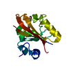
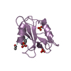
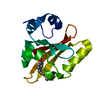
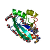
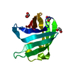
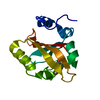
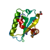
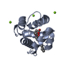
[English] 日本語
Yorodumi
- PDB-7cio: Molecular interactions of cytoplasmic region of CTLA-4 with SH2 d... -

+
Open data


ID or keywords:

Loading...

-
Basic information

Entry
Database: PDB / ID: 7cio
TitleMolecular interactions of cytoplasmic region of CTLA-4 with SH2 domains of PI3-kinase
Components
  • Cytotoxic T-lymphocyte protein 4
  • Phosphatidylinositol 3-kinase regulatory subunit alpha
KeywordsSIGNALING PROTEIN / T-CELL / CD28 / PHOSPHOPEPTIDES / PROTEIN ENGINEERING / THERMOSTABILITY
Function / homology
Function and homology information


protein complex involved in cell adhesion / negative regulation of regulatory T cell differentiation / perinuclear endoplasmic reticulum membrane / RUNX1 and FOXP3 control the development of regulatory T lymphocytes (Tregs) / regulation of toll-like receptor 4 signaling pathway / phosphatidylinositol kinase activity / phosphatidylinositol 3-kinase regulator activity / clathrin-coated endocytic vesicle / positive regulation of focal adhesion disassembly / IRS-mediated signalling ...protein complex involved in cell adhesion / negative regulation of regulatory T cell differentiation / perinuclear endoplasmic reticulum membrane / RUNX1 and FOXP3 control the development of regulatory T lymphocytes (Tregs) / regulation of toll-like receptor 4 signaling pathway / phosphatidylinositol kinase activity / phosphatidylinositol 3-kinase regulator activity / clathrin-coated endocytic vesicle / positive regulation of focal adhesion disassembly / IRS-mediated signalling / phosphatidylinositol 3-kinase activator activity / interleukin-18-mediated signaling pathway / PI3K events in ERBB4 signaling / myeloid leukocyte migration / 1-phosphatidylinositol-3-kinase regulator activity / phosphatidylinositol 3-kinase regulatory subunit binding / neurotrophin TRKA receptor binding / Activated NTRK2 signals through PI3K / Activated NTRK3 signals through PI3K / cis-Golgi network / ErbB-3 class receptor binding / RHOF GTPase cycle / kinase activator activity / transmembrane receptor protein tyrosine kinase adaptor activity / RHOD GTPase cycle / Signaling by cytosolic FGFR1 fusion mutants / positive regulation of endoplasmic reticulum unfolded protein response / enzyme-substrate adaptor activity / phosphatidylinositol 3-kinase complex, class IA / phosphatidylinositol 3-kinase complex / Nephrin family interactions / RND1 GTPase cycle / Costimulation by the CD28 family / positive regulation of leukocyte migration / MET activates PI3K/AKT signaling / RND2 GTPase cycle / PI3K/AKT activation / positive regulation of filopodium assembly / RND3 GTPase cycle / negative regulation of stress fiber assembly / growth hormone receptor signaling pathway / insulin binding / RHOV GTPase cycle / negative regulation of cell-matrix adhesion / Signaling by ALK / RHOB GTPase cycle / CTLA4 inhibitory signaling / GP1b-IX-V activation signalling / PI-3K cascade:FGFR3 / Erythropoietin activates Phosphoinositide-3-kinase (PI3K) / PI-3K cascade:FGFR2 / RHOJ GTPase cycle / PI-3K cascade:FGFR4 / RHOC GTPase cycle / negative regulation of B cell proliferation / PI-3K cascade:FGFR1 / negative regulation of osteoclast differentiation / intracellular glucose homeostasis / CD28 dependent PI3K/Akt signaling / Synthesis of PIPs at the plasma membrane / phosphatidylinositol phosphate biosynthetic process / CDC42 GTPase cycle / RHOU GTPase cycle / PI3K events in ERBB2 signaling / T cell differentiation / RET signaling / insulin receptor substrate binding / Interleukin-3, Interleukin-5 and GM-CSF signaling / RHOG GTPase cycle / extrinsic apoptotic signaling pathway via death domain receptors / PI3K Cascade / RHOA GTPase cycle / RAC2 GTPase cycle / RAC3 GTPase cycle / Role of phospholipids in phagocytosis / GAB1 signalosome / Role of LAT2/NTAL/LAB on calcium mobilization / Interleukin receptor SHC signaling / phosphatidylinositol 3-kinase binding / Signaling by PDGFRA transmembrane, juxtamembrane and kinase domain mutants / Signaling by PDGFRA extracellular domain mutants / positive regulation of lamellipodium assembly / Signaling by FGFR4 in disease / negative regulation of T cell proliferation / Signaling by FLT3 ITD and TKD mutants / Signaling by FGFR3 in disease / Tie2 Signaling / GPVI-mediated activation cascade / Signaling by FGFR2 in disease / RAC1 GTPase cycle / Signaling by FLT3 fusion proteins / FLT3 Signaling / Signaling by FGFR1 in disease / Interleukin-7 signaling / phosphotyrosine residue binding / response to endoplasmic reticulum stress / Downstream signal transduction / B cell differentiation / phosphatidylinositol 3-kinase/protein kinase B signal transduction / substrate adhesion-dependent cell spreading
Similarity search - Function
Cytotoxic T-lymphocyte antigen 4 / Cytotoxic T-lymphocyte protein 4/CD28 / Phosphatidylinositol 3-kinase regulatory subunit alpha, SH3 domain / PIK3R1, inter-SH2 domain / PI3K p85 subunit, C-terminal SH2 domain / PI3K regulatory subunit p85-related , inter-SH2 domain / PI3K p85 subunit, N-terminal SH2 domain / Phosphatidylinositol 3-kinase regulatory subunit P85 inter-SH2 domain / Rho GTPase-activating protein domain / RhoGAP domain ...Cytotoxic T-lymphocyte antigen 4 / Cytotoxic T-lymphocyte protein 4/CD28 / Phosphatidylinositol 3-kinase regulatory subunit alpha, SH3 domain / PIK3R1, inter-SH2 domain / PI3K p85 subunit, C-terminal SH2 domain / PI3K regulatory subunit p85-related , inter-SH2 domain / PI3K p85 subunit, N-terminal SH2 domain / Phosphatidylinositol 3-kinase regulatory subunit P85 inter-SH2 domain / Rho GTPase-activating protein domain / RhoGAP domain / Rho GTPase-activating proteins domain profile. / GTPase-activator protein for Rho-like GTPases / Rho GTPase activation protein / Immunoglobulin V-Type / Immunoglobulin V-set domain / SH2 domain / Src homology 2 (SH2) domain profile. / Src homology 2 domains / SH2 domain / Immunoglobulin V-set domain / Src homology 3 domains / SH2 domain superfamily / SH3-like domain superfamily / Src homology 3 (SH3) domain profile. / SH3 domain / Immunoglobulin subtype / Immunoglobulin / Immunoglobulin-like domain superfamily / Immunoglobulin-like fold
Similarity search - Domain/homology
Cytotoxic T-lymphocyte protein 4 / Phosphatidylinositol 3-kinase regulatory subunit alpha
Similarity search - Component
Biological speciesHomo sapiens (human)
MethodX-RAY DIFFRACTION / SYNCHROTRON / MOLECULAR REPLACEMENT / Resolution: 1.1 Å
AuthorsIiyama, M. / Numoto, N. / Ogawa, S. / Kuroda, M. / Morii, H. / Abe, R. / Ito, N. / Oda, M.
CitationJournal: Mol.Immunol. / Year: 2021
Title: Molecular interactions of the CTLA-4 cytoplasmic region with the phosphoinositide 3-kinase SH2 domains.
Authors: Iiyama, M. / Numoto, N. / Ogawa, S. / Kuroda, M. / Morii, H. / Abe, R. / Ito, N. / Oda, M.
History
DepositionJul 8, 2020Deposition site: PDBJ / Processing site: PDBJ
Revision 1.0Dec 9, 2020Provider: repository / Type: Initial release
Revision 1.1Mar 17, 2021Group: Database references / Category: citation
Item: _citation.journal_volume / _citation.page_first ..._citation.journal_volume / _citation.page_first / _citation.page_last / _citation.pdbx_database_id_DOI / _citation.pdbx_database_id_PubMed / _citation.year
Revision 1.2Nov 29, 2023Group: Data collection / Database references / Refinement description
Category: chem_comp_atom / chem_comp_bond ...chem_comp_atom / chem_comp_bond / database_2 / pdbx_initial_refinement_model
Item: _database_2.pdbx_DOI / _database_2.pdbx_database_accession

-
Structure visualization

Structure viewerMolecule:
MolmilJmol/JSmol

Downloads & links

-
Assembly

Deposited unit
A: Phosphatidylinositol 3-kinase regulatory subunit alpha
B: Cytotoxic T-lymphocyte protein 4


Theoretical massNumber of molelcules
Total (without water)13,1502
Polymers13,1502
Non-polymers00
Water3,045169
1


  • Idetical with deposited unit
  • defined by author&software
  • Evidence: gel filtration
TypeNameSymmetry operationNumber
identity operation1_555x,y,z1
Buried area1010 Å2
ΔGint-8 kcal/mol
Surface area6160 Å2
MethodPISA
Unit cell
Length a, b, c (Å)41.221, 41.563, 59.628
Angle α, β, γ (deg.)90.000, 90.000, 90.000
Int Tables number19
Space group name H-MP212121
Space group name HallP2ac2ab
Symmetry operation#1: x,y,z
#2: x+1/2,-y+1/2,-z
#3: -x,y+1/2,-z+1/2
#4: -x+1/2,-y,z+1/2

-
Components

#1: Protein Phosphatidylinositol 3-kinase regulatory subunit alpha / PtdIns-3-kinase regulatory subunit alpha / Phosphatidylinositol 3-kinase 85 kDa regulatory subunit ...PtdIns-3-kinase regulatory subunit alpha / Phosphatidylinositol 3-kinase 85 kDa regulatory subunit alpha / PtdIns-3-kinase regulatory subunit p85-alpha


Mass: 12178.474 Da / Num. of mol.: 1 / Mutation: C656S, C659V, C670L
Source method: isolated from a genetically manipulated source
Source: (gene. exp.) Homo sapiens (human) / Gene: PIK3R1, GRB1 / Plasmid: pGEX-4T-2 / Production host: Escherichia coli BL21(DE3) (bacteria) / References: UniProt: P27986
#2: Protein/peptide Cytotoxic T-lymphocyte protein 4 / Cytotoxic T-lymphocyte-associated antigen 4 / CTLA-4


Mass: 971.087 Da / Num. of mol.: 1 / Source method: obtained synthetically / Source: (synth.) Homo sapiens (human) / References: UniProt: P16410
#3: Water ChemComp-HOH / water


Mass: 18.015 Da / Num. of mol.: 169 / Source method: isolated from a natural source / Formula: H2O
Has ligand of interestY

-
Experimental details

-
Experiment

ExperimentMethod: X-RAY DIFFRACTION / Number of used crystals: 1

-
Sample preparation

CrystalDensity Matthews: 1.94 Å3/Da / Density % sol: 36.67 %
Crystal growTemperature: 293 K / Method: vapor diffusion, hanging drop / pH: 4.6
Details: 0.2 M ammonium acetate, 0.1 M sodium acetate trihydrate pH 4.6, 30% PEG 4000

-
Data collection

DiffractionMean temperature: 95 K / Serial crystal experiment: N
Diffraction sourceSource: SYNCHROTRON / Site: Photon Factory / Beamline: BL-17A / Wavelength: 0.98 Å
DetectorType: DECTRIS PILATUS3 S 6M / Detector: PIXEL / Date: May 18, 2019
RadiationProtocol: SINGLE WAVELENGTH / Monochromatic (M) / Laue (L): M / Scattering type: x-ray
Radiation wavelengthWavelength: 0.98 Å / Relative weight: 1
ReflectionResolution: 1.1→50 Å / Num. obs: 40740 / % possible obs: 96.3 % / Redundancy: 6.1 % / Biso Wilson estimate: 12.24 Å2 / CC1/2: 0.999 / Rsym value: 0.062 / Net I/σ(I): 15.4
Reflection shellResolution: 1.1→1.17 Å / Redundancy: 4.8 % / Mean I/σ(I) obs: 1.8 / Num. unique obs: 5178 / CC1/2: 0.689 / Rsym value: 0.884 / % possible all: 77.2

-
Processing

Software
NameVersionClassification
PHENIX1.13_2998refinement
XDSdata reduction
XDSdata scaling
PHASERphasing
RefinementMethod to determine structure: MOLECULAR REPLACEMENT
Starting model: 5AUL
Resolution: 1.1→34.1 Å / SU ML: 0.113 / Cross valid method: FREE R-VALUE / σ(F): 1.36 / Phase error: 17.8847 / Stereochemistry target values: GeoStd + Monomer Library
RfactorNum. reflection% reflection
Rfree0.1787 2037 5 %
Rwork0.1537 38688 -
obs0.1549 40725 96.25 %
Solvent computationShrinkage radii: 0.9 Å / VDW probe radii: 1.11 Å / Solvent model: FLAT BULK SOLVENT MODEL
Displacement parametersBiso mean: 17.78 Å2
Refinement stepCycle: LAST / Resolution: 1.1→34.1 Å
ProteinNucleic acidLigandSolventTotal
Num. atoms927 0 0 169 1096
Refine LS restraints
Refine-IDTypeDev idealNumber
X-RAY DIFFRACTIONf_bond_d0.0123984
X-RAY DIFFRACTIONf_angle_d1.31181346
X-RAY DIFFRACTIONf_chiral_restr0.1075148
X-RAY DIFFRACTIONf_plane_restr0.0097173
X-RAY DIFFRACTIONf_dihedral_angle_d11.832365
LS refinement shell
Resolution (Å)Rfactor RfreeNum. reflection RfreeRfactor RworkNum. reflection RworkRefine-ID% reflection obs (%)
1.1-1.130.2568870.241749X-RAY DIFFRACTION65.97
1.13-1.150.25371140.22272128X-RAY DIFFRACTION80.85
1.15-1.180.21861400.2022568X-RAY DIFFRACTION97.69
1.18-1.220.26181260.19542651X-RAY DIFFRACTION99.96
1.22-1.260.24121500.19832630X-RAY DIFFRACTION99.96
1.26-1.30.23081610.19262636X-RAY DIFFRACTION99.93
1.3-1.360.2291420.1812645X-RAY DIFFRACTION99.93
1.36-1.420.17351350.16362664X-RAY DIFFRACTION99.82
1.42-1.490.16451280.14952692X-RAY DIFFRACTION99.89
1.49-1.590.16871450.12982646X-RAY DIFFRACTION99.86
1.59-1.710.17971410.12242674X-RAY DIFFRACTION99.93
1.71-1.880.17221560.13932676X-RAY DIFFRACTION99.82
1.88-2.150.15831220.13672731X-RAY DIFFRACTION99.96
2.15-2.710.18711390.15432732X-RAY DIFFRACTION99.76
2.71-34.10.1571510.1512866X-RAY DIFFRACTION99.67

+
About Yorodumi

-
News

-
Feb 9, 2022. New format data for meta-information of EMDB entries

New format data for meta-information of EMDB entries

  • Version 3 of the EMDB header file is now the official format.
  • The previous official version 1.9 will be removed from the archive.

Related info.:EMDB header

External links:wwPDB to switch to version 3 of the EMDB data model

-
Aug 12, 2020. Covid-19 info

Covid-19 info

URL: https://pdbjlvh1.pdbj.org/emnavi/covid19.php

New page: Covid-19 featured information page in EM Navigator.

Related info.:Covid-19 info / Mar 5, 2020. Novel coronavirus structure data

+
Mar 5, 2020. Novel coronavirus structure data

Novel coronavirus structure data

Related info.:Yorodumi Speices / Aug 12, 2020. Covid-19 info

External links:COVID-19 featured content - PDBj / Molecule of the Month (242):Coronavirus Proteases

+
Jan 31, 2019. EMDB accession codes are about to change! (news from PDBe EMDB page)

EMDB accession codes are about to change! (news from PDBe EMDB page)

  • The allocation of 4 digits for EMDB accession codes will soon come to an end. Whilst these codes will remain in use, new EMDB accession codes will include an additional digit and will expand incrementally as the available range of codes is exhausted. The current 4-digit format prefixed with “EMD-” (i.e. EMD-XXXX) will advance to a 5-digit format (i.e. EMD-XXXXX), and so on. It is currently estimated that the 4-digit codes will be depleted around Spring 2019, at which point the 5-digit format will come into force.
  • The EM Navigator/Yorodumi systems omit the EMD- prefix.

Related info.:Q: What is EMD? / ID/Accession-code notation in Yorodumi/EM Navigator

External links:EMDB Accession Codes are Changing Soon! / Contact to PDBj

+
Jul 12, 2017. Major update of PDB

Major update of PDB

  • wwPDB released updated PDB data conforming to the new PDBx/mmCIF dictionary.
  • This is a major update changing the version number from 4 to 5, and with Remediation, in which all the entries are updated.
  • In this update, many items about electron microscopy experimental information are reorganized (e.g. em_software).
  • Now, EM Navigator and Yorodumi are based on the updated data.

External links:wwPDB Remediation / Enriched Model Files Conforming to OneDep Data Standards Now Available in the PDB FTP Archive

-
Yorodumi

Thousand views of thousand structures

  • Yorodumi is a browser for structure data from EMDB, PDB, SASBDB, etc.
  • This page is also the successor to EM Navigator detail page, and also detail information page/front-end page for Omokage search.
  • The word "yorodu" (or yorozu) is an old Japanese word meaning "ten thousand". "mi" (miru) is to see.

Related info.:EMDB / PDB / SASBDB / Comparison of 3 databanks / Yorodumi Search / Aug 31, 2016. New EM Navigator & Yorodumi / Yorodumi Papers / Jmol/JSmol / Function and homology information / Changes in new EM Navigator and Yorodumi

Read more