 Movie
Movie Controller


+ Open data

- Basic information
| Entry | Database: PDB / ID: 5aul | ||||||
|---|---|---|---|---|---|---|---|
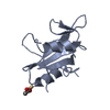
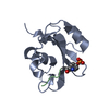
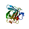
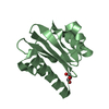
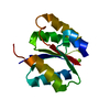
| Title | PI3K p85 C-terminal SH2 domain/CD28-derived peptide complex | ||||||
 Components Components |
| ||||||
 Keywords Keywords | SIGNALING PROTEIN / Antigens / Phosphopeptides | ||||||
| Function / homology |  Function and homology information Function and homology informationNef mediated downregulation of CD28 cell surface expression / positive regulation of inflammatory response to antigenic stimulus / protein complex involved in cell adhesion / perinuclear endoplasmic reticulum membrane / regulation of toll-like receptor 4 signaling pathway / regulation of regulatory T cell differentiation / CD4-positive, alpha-beta T cell proliferation / positive regulation of isotype switching to IgG isotypes / phosphatidylinositol kinase activity / phosphatidylinositol 3-kinase regulator activity ...Nef mediated downregulation of CD28 cell surface expression / positive regulation of inflammatory response to antigenic stimulus / protein complex involved in cell adhesion / perinuclear endoplasmic reticulum membrane / regulation of toll-like receptor 4 signaling pathway / regulation of regulatory T cell differentiation / CD4-positive, alpha-beta T cell proliferation / positive regulation of isotype switching to IgG isotypes / phosphatidylinositol kinase activity / phosphatidylinositol 3-kinase regulator activity / 1-phosphatidylinositol-3-kinase regulator activity / positive regulation of endoplasmic reticulum unfolded protein response / phosphatidylinositol 3-kinase activator activity / T follicular helper cell differentiation / IRS-mediated signalling / interleukin-18-mediated signaling pathway / negative thymic T cell selection / myeloid leukocyte migration / phosphatidylinositol 3-kinase regulatory subunit binding / PI3K events in ERBB4 signaling / neurotrophin TRKA receptor binding / regulatory T cell differentiation / positive regulation of focal adhesion disassembly / positive regulation of CD4-positive, alpha-beta T cell proliferation / cis-Golgi network / Activated NTRK2 signals through PI3K / ErbB-3 class receptor binding / transmembrane receptor protein tyrosine kinase adaptor activity / negative regulation of stress fiber assembly / Activated NTRK3 signals through PI3K / positive regulation of T cell receptor signaling pathway / phosphatidylinositol 3-kinase complex / Co-stimulation by ICOS / RHOD GTPase cycle / Signaling by cytosolic FGFR1 fusion mutants / Co-stimulation by CD28 / Nephrin family interactions / RHOF GTPase cycle / kinase activator activity / Signaling by LTK in cancer / CD28 dependent Vav1 pathway / Signaling by LTK / positive regulation of leukocyte migration / RND1 GTPase cycle / RND2 GTPase cycle / phosphatidylinositol 3-kinase complex, class IA / RND3 GTPase cycle / positive regulation of filopodium assembly / MET activates PI3K/AKT signaling / PI3K/AKT activation / growth hormone receptor signaling pathway / insulin binding / Signaling by ALK / RHOV GTPase cycle / RHOB GTPase cycle / natural killer cell mediated cytotoxicity / GP1b-IX-V activation signalling / Erythropoietin activates Phosphoinositide-3-kinase (PI3K) / PI-3K cascade:FGFR3 / PI-3K cascade:FGFR2 / PI-3K cascade:FGFR4 / PI-3K cascade:FGFR1 / RHOJ GTPase cycle / RHOC GTPase cycle / positive regulation of interleukin-4 production / negative regulation of osteoclast differentiation / phosphatidylinositol phosphate biosynthetic process / Synthesis of PIPs at the plasma membrane / RHOU GTPase cycle / humoral immune response / CDC42 GTPase cycle / RET signaling / positive regulation of interleukin-10 production / T cell differentiation / Interleukin-3, Interleukin-5 and GM-CSF signaling / PI3K events in ERBB2 signaling / RHOG GTPase cycle / immunological synapse / insulin receptor substrate binding / negative regulation of cell-matrix adhesion / PI3K Cascade / extrinsic apoptotic signaling pathway via death domain receptors / Role of LAT2/NTAL/LAB on calcium mobilization / RAC3 GTPase cycle / RHOA GTPase cycle / CD28 dependent PI3K/Akt signaling / RAC2 GTPase cycle / Interleukin receptor SHC signaling / Role of phospholipids in phagocytosis / enzyme-substrate adaptor activity / GAB1 signalosome / positive regulation of interleukin-2 production / positive regulation of viral genome replication / phosphatidylinositol 3-kinase binding / coreceptor activity / GPVI-mediated activation cascade / Signaling by PDGFRA transmembrane, juxtamembrane and kinase domain mutants / Signaling by PDGFRA extracellular domain mutants / positive regulation of lamellipodium assembly / Signaling by FGFR4 in disease Similarity search - Function | ||||||
| Biological species |  Homo sapiens (human) Homo sapiens (human) | ||||||
| Method |  X-RAY DIFFRACTION / SYNCHROTRON / MOLECULAR REPLACEMENT / Resolution: 1.1 Å X-RAY DIFFRACTION / SYNCHROTRON / MOLECULAR REPLACEMENT / Resolution: 1.1 Å | ||||||
 Authors Authors | Inaba, S. / Numoto, N. / Morii, H. / Ikura, T. / Oda, M. / Ito, N. | ||||||
 Citation Citation | Journal: J. Biol. Chem. / Year: 2017 Title: Crystal Structures and Thermodynamic Analysis Reveal Distinct Mechanisms of CD28 Phosphopeptide Binding to the Src Homology 2 (SH2) Domains of Three Adaptor Proteins Authors: Inaba, S. / Numoto, N. / Ogawa, S. / Morii, H. / Ikura, T. / Abe, R. / Ito, N. / Oda, M. | ||||||
| History |
|
- Structure visualization
| Structure viewer | Molecule: MolmilJmol/JSmol |
|---|
- Downloads & links
-Download
| PDBx/mmCIF format | 5aul.cif.gz | 78.1 KB | Display | PDBx/mmCIF format |
|---|---|---|---|---|
| PDB format | pdb5aul.ent.gz | 57.5 KB | Display | PDB format |
| PDBx/mmJSON format | 5aul.json.gz | Tree view | PDBx/mmJSON format | |
| Others |  Other downloads Other downloads |
-Validation report
| Arichive directory | https://data.pdbj.org/pub/pdb/validation_reports/au/5aulftp://data.pdbj.org/pub/pdb/validation_reports/au/5aul | HTTPS FTP |
|---|
-Related structure data
| Related structure data | 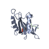 5gjhC  5gjiC  1h9oS S: Starting model for refinement C: citing same article ( |
|---|---|
| Similar structure data |


-Links
 PDBj
PDBj


- Assembly
| Deposited unit | 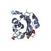
| ||||||||
|---|---|---|---|---|---|---|---|---|---|
| 1 |
| ||||||||
| Unit cell |
|
-Components
| #1: Protein | Mass: 12204.536 Da / Num. of mol.: 1 / Fragment: UNP residues 614-720 Source method: isolated from a genetically manipulated source Source: (gene. exp.) Homo sapiens (human) / Gene: PIK3R1, GRB1 / Production host:  |
|---|---|
| #2: Protein/peptide | Mass: 1038.047 Da / Num. of mol.: 1 / Fragment: UNP residues 189-196 / Source method: obtained synthetically / Source: (synth.) Homo sapiens (human) / References: UniProt: P10747 |
| #3: Chemical | ChemComp-GOL /   Mass: 92.094 Da / Num. of mol.: 1 / Source method: obtained synthetically / Formula: C3H8O3 Mass: 92.094 Da / Num. of mol.: 1 / Source method: obtained synthetically / Formula: C3H8O3 |
| #4: Water | ChemComp-HOH /  Mass: 18.015 Da / Num. of mol.: 181 / Source method: isolated from a natural source / Formula: H2O Mass: 18.015 Da / Num. of mol.: 181 / Source method: isolated from a natural source / Formula: H2O |
| Has protein modification | Y |
-Experimental details
-Experiment
| Experiment | Method: X-RAY DIFFRACTION |
|---|
- Sample preparation
| Crystal | Density Matthews: 2.06 Å3/Da / Density % sol: 40.29 % |
|---|---|
| Crystal grow | Temperature: 293 K / Method: vapor diffusion, hanging drop Details: 100 mM Sodium Acetate trihydrate, 200 mM Ammonium Acetate, 30% PEG4000 PH range: 4.6 |
-Data collection
| Diffraction | Mean temperature: 95 K |
|---|---|
| Diffraction source | Source: SYNCHROTRON / Site: Photon Factory  / Beamline: BL-17A / Wavelength: 0.98 Å / Beamline: BL-17A / Wavelength: 0.98 Å |
| Detector | Type: ADSC QUANTUM 270 / Detector: CCD / Date: Jan 27, 2014 |
| Radiation | Protocol: SINGLE WAVELENGTH / Monochromatic (M) / Laue (L): M / Scattering type: x-ray |
| Radiation wavelength | Wavelength: 0.98 Å / Relative weight: 1 |
| Reflection | Resolution: 1.1→50 Å / Num. obs: 45133 / % possible obs: 100 % / Redundancy: 8.5 % / Rmerge(I) obs: 0.068 / Net I/σ(I): 17.7 |
| Reflection shell | Resolution: 1.1→1.13 Å / Redundancy: 6.9 % / Rmerge(I) obs: 0.32 / Mean I/σ(I) obs: 6.3 / % possible all: 100 |
- Processing
| Software |
| |||||||||||||||||||||||||||||||||||||||||||||||||||||||||||||||||||||||||||||||||||||||||||||||||||||||||||||||||||||||
|---|---|---|---|---|---|---|---|---|---|---|---|---|---|---|---|---|---|---|---|---|---|---|---|---|---|---|---|---|---|---|---|---|---|---|---|---|---|---|---|---|---|---|---|---|---|---|---|---|---|---|---|---|---|---|---|---|---|---|---|---|---|---|---|---|---|---|---|---|---|---|---|---|---|---|---|---|---|---|---|---|---|---|---|---|---|---|---|---|---|---|---|---|---|---|---|---|---|---|---|---|---|---|---|---|---|---|---|---|---|---|---|---|---|---|---|---|---|---|---|---|
| Refinement | Method to determine structure: MOLECULAR REPLACEMENT Starting model: 1H9O Resolution: 1.1→34.919 Å / SU ML: 0.07 / Cross valid method: FREE R-VALUE / σ(F): 1.37 / Phase error: 13.16 / Stereochemistry target values: ML
| |||||||||||||||||||||||||||||||||||||||||||||||||||||||||||||||||||||||||||||||||||||||||||||||||||||||||||||||||||||||
| Solvent computation | Shrinkage radii: 0.9 Å / VDW probe radii: 1.11 Å / Solvent model: FLAT BULK SOLVENT MODEL | |||||||||||||||||||||||||||||||||||||||||||||||||||||||||||||||||||||||||||||||||||||||||||||||||||||||||||||||||||||||
| Refinement step | Cycle: LAST / Resolution: 1.1→34.919 Å
| |||||||||||||||||||||||||||||||||||||||||||||||||||||||||||||||||||||||||||||||||||||||||||||||||||||||||||||||||||||||
| Refine LS restraints |
| |||||||||||||||||||||||||||||||||||||||||||||||||||||||||||||||||||||||||||||||||||||||||||||||||||||||||||||||||||||||
| LS refinement shell |
|
