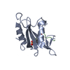
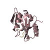
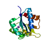
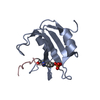
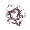
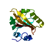
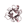
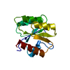
[English] 日本語
Yorodumi
- PDB-5gji: PI3K p85 N-terminal SH2 domain/CD28-derived peptide complex -

+
Open data


ID or keywords:

Loading...

-
Basic information

Entry
Database: PDB / ID: 5gji
TitlePI3K p85 N-terminal SH2 domain/CD28-derived peptide complex
Components
  • Phosphatidylinositol 3-kinase regulatory subunit alpha
  • T-cell-specific surface glycoprotein CD28
KeywordsSIGNALING PROTEIN / Antigens / Phosphopeptides
Function / homology
Function and homology information


Nef mediated downregulation of CD28 cell surface expression / regulatory T cell differentiation / positive regulation of inflammatory response to antigenic stimulus / protein complex involved in cell adhesion / regulation of regulatory T cell differentiation / perinuclear endoplasmic reticulum membrane / regulation of toll-like receptor 4 signaling pathway / CD4-positive, alpha-beta T cell proliferation / positive regulation of isotype switching to IgG isotypes / negative thymic T cell selection ...Nef mediated downregulation of CD28 cell surface expression / regulatory T cell differentiation / positive regulation of inflammatory response to antigenic stimulus / protein complex involved in cell adhesion / regulation of regulatory T cell differentiation / perinuclear endoplasmic reticulum membrane / regulation of toll-like receptor 4 signaling pathway / CD4-positive, alpha-beta T cell proliferation / positive regulation of isotype switching to IgG isotypes / negative thymic T cell selection / phosphatidylinositol kinase activity / phosphatidylinositol 3-kinase regulator activity / positive regulation of focal adhesion disassembly / IRS-mediated signalling / phosphatidylinositol 3-kinase activator activity / interleukin-18-mediated signaling pathway / PI3K events in ERBB4 signaling / myeloid leukocyte migration / positive regulation of CD4-positive, alpha-beta T cell proliferation / 1-phosphatidylinositol-3-kinase regulator activity / CD28 co-stimulation / phosphatidylinositol 3-kinase regulatory subunit binding / neurotrophin TRKA receptor binding / Activated NTRK2 signals through PI3K / Activated NTRK3 signals through PI3K / cis-Golgi network / ErbB-3 class receptor binding / kinase activator activity / RHOF GTPase cycle / transmembrane receptor protein tyrosine kinase adaptor activity / RHOD GTPase cycle / Signaling by cytosolic FGFR1 fusion mutants / positive regulation of endoplasmic reticulum unfolded protein response / enzyme-substrate adaptor activity / phosphatidylinositol 3-kinase complex, class IA / phosphatidylinositol 3-kinase complex / Nephrin family interactions / RND1 GTPase cycle / Costimulation by the CD28 family / CD28 dependent Vav1 pathway / positive regulation of leukocyte migration / MET activates PI3K/AKT signaling / RND2 GTPase cycle / PI3K/AKT activation / positive regulation of filopodium assembly / RND3 GTPase cycle / negative regulation of stress fiber assembly / growth hormone receptor signaling pathway / insulin binding / RHOV GTPase cycle / negative regulation of cell-matrix adhesion / Signaling by ALK / RHOB GTPase cycle / GP1b-IX-V activation signalling / PI-3K cascade:FGFR3 / Erythropoietin activates Phosphoinositide-3-kinase (PI3K) / positive regulation of interleukin-4 production / PI-3K cascade:FGFR2 / RHOJ GTPase cycle / PI-3K cascade:FGFR4 / RHOC GTPase cycle / PI-3K cascade:FGFR1 / negative regulation of osteoclast differentiation / intracellular glucose homeostasis / CD28 dependent PI3K/Akt signaling / Synthesis of PIPs at the plasma membrane / phosphatidylinositol phosphate biosynthetic process / positive regulation of interleukin-10 production / CDC42 GTPase cycle / RHOU GTPase cycle / humoral immune response / PI3K events in ERBB2 signaling / immunological synapse / T cell differentiation / RET signaling / insulin receptor substrate binding / Interleukin-3, Interleukin-5 and GM-CSF signaling / RHOG GTPase cycle / extrinsic apoptotic signaling pathway via death domain receptors / PI3K Cascade / RHOA GTPase cycle / RAC2 GTPase cycle / RAC3 GTPase cycle / Role of phospholipids in phagocytosis / GAB1 signalosome / Role of LAT2/NTAL/LAB on calcium mobilization / positive regulation of viral genome replication / Interleukin receptor SHC signaling / coreceptor activity / phosphatidylinositol 3-kinase binding / Signaling by PDGFRA transmembrane, juxtamembrane and kinase domain mutants / Signaling by PDGFRA extracellular domain mutants / positive regulation of lamellipodium assembly / Signaling by FGFR4 in disease / positive regulation of T cell proliferation / Signaling by FLT3 ITD and TKD mutants / Signaling by FGFR3 in disease / Tie2 Signaling / GPVI-mediated activation cascade / Signaling by FGFR2 in disease
Similarity search - Function
T cell antigen CD28 / ICOS V-set domain / Cytotoxic T-lymphocyte protein 4/CD28 / Phosphatidylinositol 3-kinase regulatory subunit alpha, SH3 domain / PIK3R1, inter-SH2 domain / PI3K p85 subunit, C-terminal SH2 domain / PI3K regulatory subunit p85-related , inter-SH2 domain / PI3K p85 subunit, N-terminal SH2 domain / Phosphatidylinositol 3-kinase regulatory subunit P85 inter-SH2 domain / Rho GTPase-activating protein domain ...T cell antigen CD28 / ICOS V-set domain / Cytotoxic T-lymphocyte protein 4/CD28 / Phosphatidylinositol 3-kinase regulatory subunit alpha, SH3 domain / PIK3R1, inter-SH2 domain / PI3K p85 subunit, C-terminal SH2 domain / PI3K regulatory subunit p85-related , inter-SH2 domain / PI3K p85 subunit, N-terminal SH2 domain / Phosphatidylinositol 3-kinase regulatory subunit P85 inter-SH2 domain / Rho GTPase-activating protein domain / RhoGAP domain / Rho GTPase-activating proteins domain profile. / GTPase-activator protein for Rho-like GTPases / Rho GTPase activation protein / SH2 domain / SHC Adaptor Protein / Immunoglobulin V-Type / SH2 domain / Src homology 2 (SH2) domain profile. / Src homology 2 domains / SH2 domain / Immunoglobulin V-set domain / Src homology 3 domains / SH2 domain superfamily / SH3-like domain superfamily / Src homology 3 (SH3) domain profile. / SH3 domain / Immunoglobulin-like domain superfamily / Immunoglobulin-like fold / 2-Layer Sandwich / Alpha Beta
Similarity search - Domain/homology
T-cell-specific surface glycoprotein CD28 / Phosphatidylinositol 3-kinase regulatory subunit alpha
Similarity search - Component
Biological speciesHomo sapiens (human)
MethodX-RAY DIFFRACTION / SYNCHROTRON / MOLECULAR REPLACEMENT / Resolution: 0.9 Å
AuthorsInaba, S. / Numoto, N. / Morii, H. / Ogawa, S. / Ikura, T. / Abe, R. / Ito, N. / Oda, M.
CitationJournal: J. Biol. Chem. / Year: 2017
Title: Crystal Structures and Thermodynamic Analysis Reveal Distinct Mechanisms of CD28 Phosphopeptide Binding to the Src Homology 2 (SH2) Domains of Three Adaptor Proteins
Authors: Inaba, S. / Numoto, N. / Ogawa, S. / Morii, H. / Ikura, T. / Abe, R. / Ito, N. / Oda, M.
History
DepositionJun 30, 2016Deposition site: PDBJ / Processing site: PDBJ
Revision 1.0Dec 14, 2016Provider: repository / Type: Initial release
Revision 1.1Feb 1, 2017Group: Database references
Revision 1.2May 10, 2017Group: Database references

-
Structure visualization

Structure viewerMolecule:
MolmilJmol/JSmol

Downloads & links

-
Assembly

Deposited unit
A: Phosphatidylinositol 3-kinase regulatory subunit alpha
B: T-cell-specific surface glycoprotein CD28
hetero molecules


Theoretical massNumber of molelcules
Total (without water)13,8516
Polymers13,4712
Non-polymers3804
Water3,423190
1


  • Idetical with deposited unit
  • defined by author&software
TypeNameSymmetry operationNumber
identity operation1_555x,y,z1
Buried area1840 Å2
ΔGint-46 kcal/mol
Surface area6660 Å2
MethodPISA
Unit cell
Length a, b, c (Å)27.590, 31.240, 32.250
Angle α, β, γ (deg.)73.28, 85.38, 71.73
Int Tables number1
Space group name H-MP1

-
Components

#1: Protein Phosphatidylinositol 3-kinase regulatory subunit alpha / PtdIns-3-kinase regulatory subunit alpha / Phosphatidylinositol 3-kinase 85 kDa regulatory subunit ...PtdIns-3-kinase regulatory subunit alpha / Phosphatidylinositol 3-kinase 85 kDa regulatory subunit alpha / PtdIns-3-kinase regulatory subunit p85-alpha


Mass: 12432.955 Da / Num. of mol.: 1 / Fragment: UNP residues 325-430 / Mutation: D330N
Source method: isolated from a genetically manipulated source
Source: (gene. exp.) Homo sapiens (human) / Gene: PIK3R1, GRB1 / Plasmid: pGEX-4T-2 / Production host: Escherichia coli BL21(DE3) (bacteria) / Strain (production host): BL21(DE3) / References: UniProt: P27986
#2: Protein/peptide T-cell-specific surface glycoprotein CD28 / TP44


Mass: 1038.047 Da / Num. of mol.: 1 / Fragment: UNP residues 189-196 / Source method: obtained synthetically / Source: (synth.) Homo sapiens (human) / References: UniProt: P10747
#3: Chemical ChemComp-SO4 / SULFATE ION


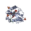
Mass: 96.063 Da / Num. of mol.: 3 / Source method: obtained synthetically / Formula: SO4
#4: Chemical ChemComp-GOL / GLYCEROL / GLYCERIN / PROPANE-1,2,3-TRIOL


Mass: 92.094 Da / Num. of mol.: 1 / Source method: obtained synthetically / Formula: C3H8O3
#5: Water ChemComp-HOH / water


Mass: 18.015 Da / Num. of mol.: 190 / Source method: isolated from a natural source / Formula: H2O

-
Experimental details

-
Experiment

ExperimentMethod: X-RAY DIFFRACTION / Number of used crystals: 1

-
Sample preparation

CrystalDensity Matthews: 1.88 Å3/Da / Density % sol: 34.45 %
Crystal growTemperature: 293 K / Method: vapor diffusion, hanging drop / pH: 4.6
Details: 100mM Sodium acetate trihydrate, 30% PEG MME 2000, 200mM Ammonium sulfate

-
Data collection

DiffractionMean temperature: 95 K
Diffraction sourceSource: SYNCHROTRON / Site: Photon Factory / Beamline: BL-17A / Wavelength: 0.98 Å
DetectorType: ADSC QUANTUM 270 / Detector: CCD / Date: May 13, 2014
RadiationProtocol: SINGLE WAVELENGTH / Monochromatic (M) / Laue (L): M / Scattering type: x-ray
Radiation wavelengthWavelength: 0.98 Å / Relative weight: 1
ReflectionResolution: 0.9→30.9 Å / Num. obs: 65076 / % possible obs: 89.5 % / Redundancy: 5.5 % / CC1/2: 0.999 / Rmerge(I) obs: 0.055 / Net I/σ(I): 18.1
Reflection shellResolution: 0.9→0.95 Å / Redundancy: 3.9 % / Rmerge(I) obs: 0.342 / Mean I/σ(I) obs: 4 / CC1/2: 0.92 / % possible all: 81.1

-
Processing

Software
NameVersionClassification
PHENIX1.9_1692refinement
XDSMarch 1, 2015data reduction
XSCALEMarch 1, 2015data scaling
MOLREPphasing
RefinementMethod to determine structure: MOLECULAR REPLACEMENT / Resolution: 0.9→30.884 Å / SU ML: 0.08 / Cross valid method: FREE R-VALUE / σ(F): 1.99 / Phase error: 12.05
RfactorNum. reflection% reflection
Rfree0.1446 3260 5.01 %
Rwork0.1259 --
obs0.1268 65076 89.68 %
Solvent computationShrinkage radii: 0.9 Å / VDW probe radii: 1.11 Å
Refinement stepCycle: LAST / Resolution: 0.9→30.884 Å
ProteinNucleic acidLigandSolventTotal
Num. atoms942 0 21 190 1153
Refine LS restraints
Refine-IDTypeDev idealNumber
X-RAY DIFFRACTIONf_bond_d0.0071120
X-RAY DIFFRACTIONf_angle_d1.3871534
X-RAY DIFFRACTIONf_dihedral_angle_d12.605435
X-RAY DIFFRACTIONf_chiral_restr0.058157
X-RAY DIFFRACTIONf_plane_restr0.007200
LS refinement shell
Resolution (Å)Rfactor RfreeNum. reflection RfreeRfactor RworkNum. reflection RworkRefine-ID% reflection obs (%)
0.9003-0.91380.23321220.23872229X-RAY DIFFRACTION75
0.9138-0.92810.21431290.19352535X-RAY DIFFRACTION85
0.9281-0.94330.18691360.15692551X-RAY DIFFRACTION84
0.9433-0.95950.14081150.13662568X-RAY DIFFRACTION86
0.9595-0.9770.1511370.12842564X-RAY DIFFRACTION85
0.977-0.99580.1561410.1222583X-RAY DIFFRACTION87
0.9958-1.01610.13181400.11042606X-RAY DIFFRACTION87
1.0161-1.03820.1091480.10512581X-RAY DIFFRACTION87
1.0382-1.06240.11591560.0972629X-RAY DIFFRACTION88
1.0624-1.08890.1041460.09472616X-RAY DIFFRACTION88
1.0889-1.11840.1131520.09232691X-RAY DIFFRACTION89
1.1184-1.15130.10131500.08842713X-RAY DIFFRACTION90
1.1513-1.18840.11071510.08892658X-RAY DIFFRACTION90
1.1884-1.23090.10631270.09222749X-RAY DIFFRACTION91
1.2309-1.28020.10821270.09732756X-RAY DIFFRACTION92
1.2802-1.33850.11331390.10332772X-RAY DIFFRACTION92
1.3385-1.4090.11951430.10452800X-RAY DIFFRACTION93
1.409-1.49730.10821590.10652798X-RAY DIFFRACTION94
1.4973-1.61290.13691420.11342825X-RAY DIFFRACTION95
1.6129-1.77520.13431320.132865X-RAY DIFFRACTION95
1.7752-2.0320.15061500.14132904X-RAY DIFFRACTION96
2.032-2.560.17231530.14642913X-RAY DIFFRACTION97
2.56-30.90230.18981650.15222910X-RAY DIFFRACTION97

+
About Yorodumi

-
News

-
Feb 9, 2022. New format data for meta-information of EMDB entries

New format data for meta-information of EMDB entries

  • Version 3 of the EMDB header file is now the official format.
  • The previous official version 1.9 will be removed from the archive.

Related info.:EMDB header

External links:wwPDB to switch to version 3 of the EMDB data model

-
Aug 12, 2020. Covid-19 info

Covid-19 info

URL: https://pdbjlvh1.pdbj.org/emnavi/covid19.php

New page: Covid-19 featured information page in EM Navigator.

Related info.:Covid-19 info / Mar 5, 2020. Novel coronavirus structure data

+
Mar 5, 2020. Novel coronavirus structure data

Novel coronavirus structure data

Related info.:Yorodumi Speices / Aug 12, 2020. Covid-19 info

External links:COVID-19 featured content - PDBj / Molecule of the Month (242):Coronavirus Proteases

+
Jan 31, 2019. EMDB accession codes are about to change! (news from PDBe EMDB page)

EMDB accession codes are about to change! (news from PDBe EMDB page)

  • The allocation of 4 digits for EMDB accession codes will soon come to an end. Whilst these codes will remain in use, new EMDB accession codes will include an additional digit and will expand incrementally as the available range of codes is exhausted. The current 4-digit format prefixed with “EMD-” (i.e. EMD-XXXX) will advance to a 5-digit format (i.e. EMD-XXXXX), and so on. It is currently estimated that the 4-digit codes will be depleted around Spring 2019, at which point the 5-digit format will come into force.
  • The EM Navigator/Yorodumi systems omit the EMD- prefix.

Related info.:Q: What is EMD? / ID/Accession-code notation in Yorodumi/EM Navigator

External links:EMDB Accession Codes are Changing Soon! / Contact to PDBj

+
Jul 12, 2017. Major update of PDB

Major update of PDB

  • wwPDB released updated PDB data conforming to the new PDBx/mmCIF dictionary.
  • This is a major update changing the version number from 4 to 5, and with Remediation, in which all the entries are updated.
  • In this update, many items about electron microscopy experimental information are reorganized (e.g. em_software).
  • Now, EM Navigator and Yorodumi are based on the updated data.

External links:wwPDB Remediation / Enriched Model Files Conforming to OneDep Data Standards Now Available in the PDB FTP Archive

-
Yorodumi

Thousand views of thousand structures

  • Yorodumi is a browser for structure data from EMDB, PDB, SASBDB, etc.
  • This page is also the successor to EM Navigator detail page, and also detail information page/front-end page for Omokage search.
  • The word "yorodu" (or yorozu) is an old Japanese word meaning "ten thousand". "mi" (miru) is to see.

Related info.:EMDB / PDB / SASBDB / Comparison of 3 databanks / Yorodumi Search / Aug 31, 2016. New EM Navigator & Yorodumi / Yorodumi Papers / Jmol/JSmol / Function and homology information / Changes in new EM Navigator and Yorodumi

Read more