 Movie
Movie Controller

[English] 日本語
 Yorodumi
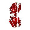
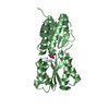
- PDB-1mvo: Crystal structure of the PhoP receiver domain from Bacillus subtilis -
+ Open data

- Basic information
| Entry | Database: PDB / ID: 1mvo | ||||||
|---|---|---|---|---|---|---|---|
| Title | Crystal structure of the PhoP receiver domain from Bacillus subtilis | ||||||
 Components Components | PhoP response regulator | ||||||
 Keywords Keywords | TRANSCRIPTION / PHOSPHATE REGULON / TRANSCRIPTIONAL REGULATORY PROTEIN / ALPHA/BETA DOUBLY WOUND FOLD / RESPONSE REGULATOR / PHOSPHORYLATION / ASYMMETRIC INTERFACE / TANDEM ASSOCIATION / Structural Proteomics in Europe / SPINE / Structural Genomics | ||||||
| Function / homology |  Function and homology information Function and homology informationphosphate ion transport / phosphorelay response regulator activity / protein-DNA complex / transcription cis-regulatory region binding / regulation of DNA-templated transcription / cytosol Similarity search - Function | ||||||
| Biological species |  | ||||||
| Method |  X-RAY DIFFRACTION / SYNCHROTRON / MOLECULAR REPLACEMENT / Resolution: 1.6 Å X-RAY DIFFRACTION / SYNCHROTRON / MOLECULAR REPLACEMENT / Resolution: 1.6 Å | ||||||
 Authors Authors | Birck, C. / Chen, Y. / Hulett, F.M. / Samama, J.P. / Structural Proteomics in Europe (SPINE) | ||||||
 Citation Citation | Journal: J.BACTERIOL. / Year: 2003 Title: The Crystal Structure of the Phosphorylation Domain in PhoP Reveals a Functional Tandem Association Mediated by an Asymmetric Interface Authors: Birck, C. / Chen, Y. / Hulett, F.M. / Samama, J.P. #1: Journal: J.BACTERIOL. / Year: 2003Title: Residue R113 Is Essential for PhoP Dimerization and Function: a Residue Buried in the Asymmetric PhoP Dimer Interface Determined in the PhoPN Three-Dimensional Crystal Structure Authors: Chen, Y. / Birck, C. / Samama, J.P. / Hulett, F.M. | ||||||
| History |
|
- Structure visualization
| Structure viewer | Molecule: MolmilJmol/JSmol |
|---|
- Downloads & links
-Download
| PDBx/mmCIF format | 1mvo.cif.gz | 41.8 KB | Display | PDBx/mmCIF format |
|---|---|---|---|---|
| PDB format | pdb1mvo.ent.gz | 28.3 KB | Display | PDB format |
| PDBx/mmJSON format | 1mvo.json.gz | Tree view | PDBx/mmJSON format | |
| Others |  Other downloads Other downloads |
-Validation report
| Arichive directory | https://data.pdbj.org/pub/pdb/validation_reports/mv/1mvoftp://data.pdbj.org/pub/pdb/validation_reports/mv/1mvo | HTTPS FTP |
|---|
-Related structure data
| Similar structure data | |
|---|---|
| Other databases |


-Links
 PDBj
PDBj

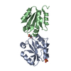
- Assembly
| Deposited unit | 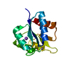
| ||||||||
|---|---|---|---|---|---|---|---|---|---|
| 1 | 
| ||||||||
| Unit cell |
| ||||||||
| Details | The biological assembly is a dimer generated from the monomer in the asymmetric unit by the operation : x+1/2, -y+3/2, -z+3/4 |
-Components
| #1: Protein | Mass: 15546.987 Da / Num. of mol.: 1 / Fragment: N-terminal domain Source method: isolated from a genetically manipulated source Source: (gene. exp.) | ||
|---|---|---|---|
| #2: Chemical | ChemComp-MN /   Mass: 54.938 Da / Num. of mol.: 1 / Source method: obtained synthetically / Formula: Mn Mass: 54.938 Da / Num. of mol.: 1 / Source method: obtained synthetically / Formula: Mn | ||
| #3: Chemical |   Mass: 22.990 Da / Num. of mol.: 2 / Source method: obtained synthetically / Formula: Na Mass: 22.990 Da / Num. of mol.: 2 / Source method: obtained synthetically / Formula: Na#4: Water | ChemComp-HOH / |  Mass: 18.015 Da / Num. of mol.: 174 / Source method: isolated from a natural source / Formula: H2O Mass: 18.015 Da / Num. of mol.: 174 / Source method: isolated from a natural source / Formula: H2O |
-Experimental details
-Experiment
| Experiment | Method: X-RAY DIFFRACTION / Number of used crystals: 1 |
|---|
- Sample preparation
| Crystal | Density Matthews: 2.26 Å3/Da / Density % sol: 45.65 % | ||||||||||||||||||||||||||||||||||||||||||||||||||||||||
|---|---|---|---|---|---|---|---|---|---|---|---|---|---|---|---|---|---|---|---|---|---|---|---|---|---|---|---|---|---|---|---|---|---|---|---|---|---|---|---|---|---|---|---|---|---|---|---|---|---|---|---|---|---|---|---|---|---|
| Crystal grow | Temperature: 277 K / Method: vapor diffusion, hanging drop / pH: 5.8 Details: PEG 10000, sodium citrate, PEG MME 550, pH 5.8, VAPOR DIFFUSION, HANGING DROP, temperature 277K | ||||||||||||||||||||||||||||||||||||||||||||||||||||||||
| Crystal grow | *PLUS Temperature: 4 ℃ / pH: 7.8 | ||||||||||||||||||||||||||||||||||||||||||||||||||||||||
| Components of the solutions | *PLUS
|
-Data collection
| Diffraction | Mean temperature: 100 K |
|---|---|
| Diffraction source | Source: SYNCHROTRON / Site: ESRF  / Beamline: ID14-1 / Wavelength: 0.934 Å / Beamline: ID14-1 / Wavelength: 0.934 Å |
| Detector | Type: MARRESEARCH / Detector: CCD / Date: Oct 4, 2000 |
| Radiation | Monochromator: SAGITALLY FOCUSED Ge(220) / Protocol: SINGLE WAVELENGTH / Monochromatic (M) / Laue (L): M / Scattering type: x-ray |
| Radiation wavelength | Wavelength: 0.934 Å / Relative weight: 1 |
| Reflection | Resolution: 1.6→27 Å / Num. all: 18122 / Num. obs: 18122 / % possible obs: 91.6 % / Observed criterion σ(F): 0 / Observed criterion σ(I): 0 / Redundancy: 6.1 % / Biso Wilson estimate: 19.8 Å2 / Rmerge(I) obs: 0.091 / Rsym value: 0.091 / Net I/σ(I): 5.2 |
| Reflection shell | Resolution: 1.6→1.69 Å / Redundancy: 4.3 % / Rmerge(I) obs: 0.187 / Mean I/σ(I) obs: 3.5 / Num. unique all: 2775 / Rsym value: 0.187 / % possible all: 92 |
| Reflection | *PLUS Lowest resolution: 27.1 Å / Num. obs: 18249 / % possible obs: 92 % / Num. measured all: 224913 |
| Reflection shell | *PLUS % possible obs: 92 % |
- Processing
| Software |
| |||||||||||||||||||||||||
|---|---|---|---|---|---|---|---|---|---|---|---|---|---|---|---|---|---|---|---|---|---|---|---|---|---|---|
| Refinement | Method to determine structure: MOLECULAR REPLACEMENT Starting model: Metal-free PhoPN structure solved by MAD Resolution: 1.6→27 Å / Isotropic thermal model: anisotropic / Cross valid method: THROUGHOUT / σ(F): 0 / Stereochemistry target values: Engh & Huber Details: Used weighted full matrix maximum likelihood procedure
| |||||||||||||||||||||||||
| Displacement parameters | Biso mean: 17.523 Å2
| |||||||||||||||||||||||||
| Refinement step | Cycle: LAST / Resolution: 1.6→27 Å
| |||||||||||||||||||||||||
| Refine LS restraints |
| |||||||||||||||||||||||||
| LS refinement shell | Resolution: 1.6→1.66 Å
| |||||||||||||||||||||||||
| Refinement | *PLUS Lowest resolution: 27.1 Å / Num. reflection obs: 17225 / Rfactor Rfree: 0.233 | |||||||||||||||||||||||||
| Solvent computation | *PLUS | |||||||||||||||||||||||||
| Displacement parameters | *PLUS |
