 Movie
Movie Controller


+ Open data

- Basic information
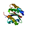
| Entry | Database: PDB / ID: 2zp2 | ||||||
|---|---|---|---|---|---|---|---|
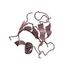
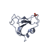
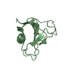
| Title | C-terminal domain of KipI from Bacillus subtilis | ||||||
 Components Components | Kinase A inhibitor | ||||||
 Keywords Keywords | TRANSFERASE INHIBITOR / kipI / histidine kinase inhibitor / ATP-binding / Nucleotide-binding / Protein kinase inhibitor / Sporulation | ||||||
| Function / homology |  Function and homology information Function and homology information5-oxoprolinase (ATP-hydrolysing) / 5-oxoprolinase (ATP-hydrolyzing) activity / sporulation resulting in formation of a cellular spore / protein kinase inhibitor activity / ATP binding Similarity search - Function | ||||||
| Biological species |  | ||||||
| Method |  X-RAY DIFFRACTION / MOLECULAR REPLACEMENT / Resolution: 3.01 Å X-RAY DIFFRACTION / MOLECULAR REPLACEMENT / Resolution: 3.01 Å | ||||||
 Authors Authors | Langley, D.B. / Jacques, D.A. | ||||||
 Citation Citation | Journal: J.Mol.Biol. / Year: 2008 Title: Histidine kinase regulation by a cyclophilin-like inhibitor Authors: Jacques, D.A. / Langley, D.B. / Jeffries, C.M. / Cunningham, K.A. / Burkholder, W.F. / Guss, J.M. / Trewhella, J. | ||||||
| History |
|
- Structure visualization
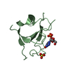
| Structure viewer | Molecule: MolmilJmol/JSmol |
|---|
- Downloads & links
-Download
| PDBx/mmCIF format | 2zp2.cif.gz | 55 KB | Display | PDBx/mmCIF format |
|---|---|---|---|---|
| PDB format | pdb2zp2.ent.gz | 38.4 KB | Display | PDB format |
| PDBx/mmJSON format | 2zp2.json.gz | Tree view | PDBx/mmJSON format | |
| Others |  Other downloads Other downloads |
-Validation report
| Arichive directory | https://data.pdbj.org/pub/pdb/validation_reports/zp/2zp2ftp://data.pdbj.org/pub/pdb/validation_reports/zp/2zp2 | HTTPS FTP |
|---|
-Related structure data
| Related structure data |  2phcS S: Starting model for refinement |
|---|---|
| Similar structure data |


-Links
 PDBj
PDBj

- Assembly
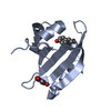
| Deposited unit | 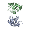
| ||||||||
|---|---|---|---|---|---|---|---|---|---|
| 1 | 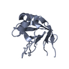
| ||||||||
| 2 | 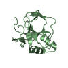
| ||||||||
| Unit cell |
|
-Components
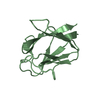
| #1: Protein | Mass: 15341.532 Da / Num. of mol.: 2 / Fragment: C-terminal domain, UNP residues 100-240 Source method: isolated from a genetically manipulated source Source: (gene. exp.) |
|---|
-Experimental details
-Experiment
| Experiment | Method: X-RAY DIFFRACTION / Number of used crystals: 1 |
|---|
- Sample preparation
| Crystal | Density Matthews: 2.61 Å3/Da / Density % sol: 52.82 % |
|---|---|
| Crystal grow | Temperature: 293 K / Method: vapor diffusion, hanging drop / pH: 6.9 Details: 15% PEG 8000, 20% glycerol, 40mM potassium phosphate, pH 6.9, VAPOR DIFFUSION, HANGING DROP, temperature 293K |
-Data collection
| Diffraction | Mean temperature: 100 K |
|---|---|
| Diffraction source | Source: ROTATING ANODE / Type: RIGAKU RU200 / Wavelength: 1.5418 Å |
| Detector | Type: MAR scanner 345 mm plate / Detector: IMAGE PLATE / Date: May 15, 2008 / Details: Osmic mirror optics |
| Radiation | Monochromator: Ni filter / Protocol: SINGLE WAVELENGTH / Monochromatic (M) / Laue (L): M / Scattering type: x-ray |
| Radiation wavelength | Wavelength: 1.5418 Å / Relative weight: 1 |
| Reflection | Resolution: 3→17 Å / Num. all: 6751 / Num. obs: 6738 / % possible obs: 99.8 % / Observed criterion σ(F): -3 / Observed criterion σ(I): -3 / Redundancy: 6 % / Biso Wilson estimate: 78 Å2 / Rmerge(I) obs: 0.14 / Net I/σ(I): 6.7 |
| Reflection shell | Resolution: 3→3.11 Å / Redundancy: 4.6 % / Rmerge(I) obs: 0.66 / Mean I/σ(I) obs: 1.5 / Num. unique all: 629 / % possible all: 98.4 |
- Processing
| Software |
| ||||||||||||||||||||||||||||||||||||||||||||||||||||||||||||||||||||||||||||||||||||||||||||||||||||||||||||||||||||||||||||||||||
|---|---|---|---|---|---|---|---|---|---|---|---|---|---|---|---|---|---|---|---|---|---|---|---|---|---|---|---|---|---|---|---|---|---|---|---|---|---|---|---|---|---|---|---|---|---|---|---|---|---|---|---|---|---|---|---|---|---|---|---|---|---|---|---|---|---|---|---|---|---|---|---|---|---|---|---|---|---|---|---|---|---|---|---|---|---|---|---|---|---|---|---|---|---|---|---|---|---|---|---|---|---|---|---|---|---|---|---|---|---|---|---|---|---|---|---|---|---|---|---|---|---|---|---|---|---|---|---|---|---|---|---|
| Refinement | Method to determine structure: MOLECULAR REPLACEMENT Starting model: PDB entry 2PHC Resolution: 3.01→16.88 Å / Cor.coef. Fo:Fc: 0.872 / Cor.coef. Fo:Fc free: 0.788 / SU B: 21.564 / SU ML: 0.415 / Isotropic thermal model: Isotropic / Cross valid method: THROUGHOUT / σ(F): 0 / ESU R Free: 0.533 / Stereochemistry target values: MAXIMUM LIKELIHOOD Details: HYDROGENS HAVE BEEN ADDED IN THE RIDING POSITIONS but are not displayed in the PDB
| ||||||||||||||||||||||||||||||||||||||||||||||||||||||||||||||||||||||||||||||||||||||||||||||||||||||||||||||||||||||||||||||||||
| Solvent computation | Ion probe radii: 0.8 Å / Shrinkage radii: 0.8 Å / VDW probe radii: 1.2 Å / Solvent model: MASK | ||||||||||||||||||||||||||||||||||||||||||||||||||||||||||||||||||||||||||||||||||||||||||||||||||||||||||||||||||||||||||||||||||
| Displacement parameters | Biso mean: 37.673 Å2
| ||||||||||||||||||||||||||||||||||||||||||||||||||||||||||||||||||||||||||||||||||||||||||||||||||||||||||||||||||||||||||||||||||
| Refinement step | Cycle: LAST / Resolution: 3.01→16.88 Å
| ||||||||||||||||||||||||||||||||||||||||||||||||||||||||||||||||||||||||||||||||||||||||||||||||||||||||||||||||||||||||||||||||||
| Refine LS restraints |
| ||||||||||||||||||||||||||||||||||||||||||||||||||||||||||||||||||||||||||||||||||||||||||||||||||||||||||||||||||||||||||||||||||
| LS refinement shell | Resolution: 3.007→3.082 Å / Total num. of bins used: 20
|
