 Movie
Movie Controller

[English] 日本語
 Yorodumi
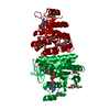
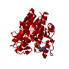
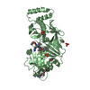
- PDB-1oh9: Acetylglutamate kinase from Escherichia coli complexed with MgADP... -
+ Open data

- Basic information
| Entry | Database: PDB / ID: 1oh9 | ||||||
|---|---|---|---|---|---|---|---|
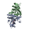
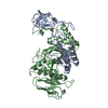
| Title | Acetylglutamate kinase from Escherichia coli complexed with MgADP, N-acetyl-L-glutamate and the transition-state mimic AlF4- | ||||||
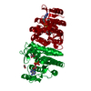
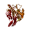
 Components Components | ACETYLGLUTAMATE KINASE | ||||||
 Keywords Keywords | KINASE / N-ACETYL-L-GLUTAMATE KINASE / AMINO ACID KINASE / PHOSPHORYL N-ACETYL-L-GLUTAMATE 5-PHOSPHOTRANSFERASE / NAG KINASE / GROUP TRANSFER / ARGININE METABOLISM | ||||||
| Function / homology |  Function and homology information Function and homology informationacetylglutamate kinase / acetylglutamate kinase activity / : / L-arginine biosynthetic process / DNA damage response / ATP binding / cytoplasm Similarity search - Function | ||||||
| Biological species |  | ||||||
| Method |  X-RAY DIFFRACTION / SYNCHROTRON / MOLECULAR REPLACEMENT / Resolution: 1.91 Å X-RAY DIFFRACTION / SYNCHROTRON / MOLECULAR REPLACEMENT / Resolution: 1.91 Å | ||||||
 Authors Authors | Gil-Ortiz, F. / Ramon-Maiques, S. / Fita, I. / Rubio, V. | ||||||
 Citation Citation | Journal: J.Mol.Biol. / Year: 2003 Title: The Course of Phosphorus in the Reaction of N-Acetyl-L-Glutamate Kinase, Determined from the Structures of Crystalline Complexes, Including a Complex with an Alf(4)(-) Transition State Mimic Authors: Gil-Ortiz, F. / Ramon-Maiques, S. / Fita, I. / Rubio, V. | ||||||
| History |
|
- Structure visualization
| Structure viewer | Molecule: MolmilJmol/JSmol |
|---|
- Downloads & links
-Download
| PDBx/mmCIF format | 1oh9.cif.gz | 69 KB | Display | PDBx/mmCIF format |
|---|---|---|---|---|
| PDB format | pdb1oh9.ent.gz | 48.9 KB | Display | PDB format |
| PDBx/mmJSON format | 1oh9.json.gz | Tree view | PDBx/mmJSON format | |
| Others |  Other downloads Other downloads |
-Validation report
| Arichive directory | https://data.pdbj.org/pub/pdb/validation_reports/oh/1oh9ftp://data.pdbj.org/pub/pdb/validation_reports/oh/1oh9 | HTTPS FTP |
|---|
-Related structure data
| Related structure data | 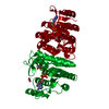 1ohaC  1ohbC 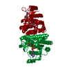 1gs5S S: Starting model for refinement C: citing same article ( |
|---|---|
| Similar structure data |


-Links
 PDBj
PDBj


- Assembly
| Deposited unit | 
| ||||||||
|---|---|---|---|---|---|---|---|---|---|
| 1 | 
| ||||||||
| Unit cell |
| ||||||||
| Components on special symmetry positions |
|
-Components
-Protein , 1 types, 1 molecules A
| #1: Protein | Mass: 27186.492 Da / Num. of mol.: 1 Source method: isolated from a genetically manipulated source Source: (gene. exp.) References: UniProt: P11445, UniProt: P0A6C8*PLUS, acetylglutamate kinase |
|---|
-Non-polymers , 5 types, 214 molecules 
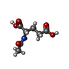


| #2: Chemical | ChemComp-ADP /  Mass: 427.201 Da / Num. of mol.: 1 / Source method: obtained synthetically / Formula: C10H15N5O10P2 / Comment: ADP, energy-carrying molecule*YM Mass: 427.201 Da / Num. of mol.: 1 / Source method: obtained synthetically / Formula: C10H15N5O10P2 / Comment: ADP, energy-carrying molecule*YM |
|---|---|
| #3: Chemical | ChemComp-NLG /  Mass: 189.166 Da / Num. of mol.: 1 / Source method: obtained synthetically / Formula: C7H11NO5 Mass: 189.166 Da / Num. of mol.: 1 / Source method: obtained synthetically / Formula: C7H11NO5 |
| #4: Chemical | ChemComp-ALF /  Mass: 102.975 Da / Num. of mol.: 1 / Source method: obtained synthetically / Formula: AlF4 Mass: 102.975 Da / Num. of mol.: 1 / Source method: obtained synthetically / Formula: AlF4 |
| #5: Chemical | ChemComp-MG /  Mass: 24.305 Da / Num. of mol.: 1 / Source method: obtained synthetically / Formula: Mg Mass: 24.305 Da / Num. of mol.: 1 / Source method: obtained synthetically / Formula: Mg |
| #6: Water | ChemComp-HOH / Mass: 18.015 Da / Num. of mol.: 210 / Source method: isolated from a natural source / Formula: H2O |
-Experimental details
-Experiment
| Experiment | Method: X-RAY DIFFRACTION / Number of used crystals: 1 |
|---|
- Sample preparation
| Crystal | Density Matthews: 2.1 Å3/Da / Density % sol: 41 % | ||||||||||||||||||||||||||||||
|---|---|---|---|---|---|---|---|---|---|---|---|---|---|---|---|---|---|---|---|---|---|---|---|---|---|---|---|---|---|---|---|
| Crystal grow | pH: 4.6 Details: 27-29% POLYETHYLENE GLYCOL MONOMETHYL ETHER 2K, SODIUM ACETATE 0.1M PH 4.6 AMMONIUM CITRATE 0.25-0.4 M | ||||||||||||||||||||||||||||||
| Crystal grow | *PLUS pH: 4.6 / Method: vapor diffusion | ||||||||||||||||||||||||||||||
| Components of the solutions | *PLUS
|
-Data collection
| Diffraction | Mean temperature: 100 K |
|---|---|
| Diffraction source | Source: SYNCHROTRON / Site: EMBL/DESY, HAMBURG  / Beamline: BW7B / Wavelength: 0.8424 / Beamline: BW7B / Wavelength: 0.8424 |
| Detector | Type: MARRESEARCH / Detector: CCD / Date: Apr 4, 2000 |
| Radiation | Protocol: SINGLE WAVELENGTH / Monochromatic (M) / Laue (L): M / Scattering type: x-ray |
| Radiation wavelength | Wavelength: 0.8424 Å / Relative weight: 1 |
| Reflection | Resolution: 1.91→30.02 Å / Num. obs: 18025 / % possible obs: 99 % / Observed criterion σ(I): 0 / Redundancy: 5 % / Biso Wilson estimate: 11.4 Å2 / Rmerge(I) obs: 0.037 / Net I/σ(I): 14.9 |
| Reflection shell | Resolution: 1.91→2.02 Å / Redundancy: 3.8 % / Rmerge(I) obs: 0.144 / Mean I/σ(I) obs: 5.3 / % possible all: 95 |
| Reflection | *PLUS Highest resolution: 1.91 Å / Lowest resolution: 30.02 Å / Num. measured all: 89855 / Rmerge(I) obs: 0.037 |
| Reflection shell | *PLUS % possible obs: 95 % / Rmerge(I) obs: 0.144 / Mean I/σ(I) obs: 5.3 |
- Processing
| Software |
| ||||||||||||||||||||||||||||||||||||||||||||||||||||||||||||||||||||||||||||||||
|---|---|---|---|---|---|---|---|---|---|---|---|---|---|---|---|---|---|---|---|---|---|---|---|---|---|---|---|---|---|---|---|---|---|---|---|---|---|---|---|---|---|---|---|---|---|---|---|---|---|---|---|---|---|---|---|---|---|---|---|---|---|---|---|---|---|---|---|---|---|---|---|---|---|---|---|---|---|---|---|---|---|
| Refinement | Method to determine structure: MOLECULAR REPLACEMENT Starting model: PDB ENTRY 1GS5 Resolution: 1.91→30 Å / Rfactor Rfree error: 0.0078 / Data cutoff high absF: 10000 / Isotropic thermal model: RESTRAINED / Cross valid method: THROUGHOUT / σ(F): 0 Details: ISOMORPHOUS CRYSTALS WITH 1GS5 ALLOWED A PURE RIGID-BODY APPROACH
| ||||||||||||||||||||||||||||||||||||||||||||||||||||||||||||||||||||||||||||||||
| Solvent computation | Solvent model: FLAT MODEL / Bsol: 52.5073 Å2 / ksol: 0.373379 e/Å3 | ||||||||||||||||||||||||||||||||||||||||||||||||||||||||||||||||||||||||||||||||
| Displacement parameters | Biso mean: 22 Å2
| ||||||||||||||||||||||||||||||||||||||||||||||||||||||||||||||||||||||||||||||||
| Refine analyze |
| ||||||||||||||||||||||||||||||||||||||||||||||||||||||||||||||||||||||||||||||||
| Refinement step | Cycle: LAST / Resolution: 1.91→30 Å
| ||||||||||||||||||||||||||||||||||||||||||||||||||||||||||||||||||||||||||||||||
| Refine LS restraints |
| ||||||||||||||||||||||||||||||||||||||||||||||||||||||||||||||||||||||||||||||||
| LS refinement shell | Resolution: 1.91→2.03 Å / Rfactor Rfree error: 0.019 / Total num. of bins used: 6
| ||||||||||||||||||||||||||||||||||||||||||||||||||||||||||||||||||||||||||||||||
| Refinement | *PLUS Num. reflection obs: 17143 | ||||||||||||||||||||||||||||||||||||||||||||||||||||||||||||||||||||||||||||||||
| Solvent computation | *PLUS | ||||||||||||||||||||||||||||||||||||||||||||||||||||||||||||||||||||||||||||||||
| Displacement parameters | *PLUS | ||||||||||||||||||||||||||||||||||||||||||||||||||||||||||||||||||||||||||||||||
| Refine LS restraints | *PLUS
|
