 Movie
Movie Controller

[English] 日本語
 Yorodumi
- PDB-1gs5: N-acetyl-L-glutamate kinase from Escherichia coli complexed with ... -
+ Open data

- Basic information
| Entry | Database: PDB / ID: 1gs5 | ||||||
|---|---|---|---|---|---|---|---|
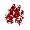
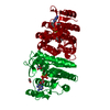
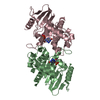
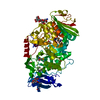
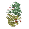
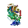
| Title | N-acetyl-L-glutamate kinase from Escherichia coli complexed with its substrate N-acetylglutamate and its substrate analog AMPPNP | ||||||
 Components Components | ACETYLGLUTAMATE KINASE | ||||||
 Keywords Keywords | TRANSFERASE / CARBAMATE KINASE / AMINO ACID KINASE / ARGININE BIOSYNTHESIS / PHOSPHORYL GROUP TRANSFER | ||||||
| Function / homology |  Function and homology information Function and homology informationacetylglutamate kinase / acetylglutamate kinase activity / : / L-arginine biosynthetic process / DNA damage response / ATP binding / cytoplasm Similarity search - Function | ||||||
| Biological species |  | ||||||
| Method |  X-RAY DIFFRACTION / SYNCHROTRON / MOLECULAR REPLACEMENT / Resolution: 1.5 Å X-RAY DIFFRACTION / SYNCHROTRON / MOLECULAR REPLACEMENT / Resolution: 1.5 Å | ||||||
 Authors Authors | Ramon-Maiques, S. / Marina, A. / Gil-Ortiz, F. / Fita, I. / Rubio, V. | ||||||
 Citation Citation | Journal: Structure / Year: 2002 Title: Structure of Acetylglutamate Kinase, a Key Enzyme for Arginine Biosynthesis and a Prototype for the Amino Acid Kinase Enzyme Family, During Catalysis Authors: Ramon-Maiques, S. / Marina, A. / Gil-Ortiz, F. / Fita, I. / Rubio, V. | ||||||
| History |
|
- Structure visualization
| Structure viewer | Molecule: MolmilJmol/JSmol |
|---|
- Downloads & links
-Download
| PDBx/mmCIF format | 1gs5.cif.gz | 68.7 KB | Display | PDBx/mmCIF format |
|---|---|---|---|---|
| PDB format | pdb1gs5.ent.gz | 49.2 KB | Display | PDB format |
| PDBx/mmJSON format | 1gs5.json.gz | Tree view | PDBx/mmJSON format | |
| Others |  Other downloads Other downloads |
-Validation report
| Arichive directory | https://data.pdbj.org/pub/pdb/validation_reports/gs/1gs5ftp://data.pdbj.org/pub/pdb/validation_reports/gs/1gs5 | HTTPS FTP |
|---|
-Related structure data
| Related structure data | 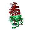 1gsjSC S: Starting model for refinement C: citing same article ( |
|---|---|
| Similar structure data |


-Links
 PDBj
PDBj


- Assembly
| Deposited unit | 
| ||||||||
|---|---|---|---|---|---|---|---|---|---|
| 1 | 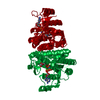
| ||||||||
| Unit cell |
| ||||||||
| Components on special symmetry positions |
|
-Components
| #1: Protein | Mass: 27186.492 Da / Num. of mol.: 1 Source method: isolated from a genetically manipulated source Source: (gene. exp.) References: UniProt: P11445, UniProt: P0A6C8*PLUS, acetylglutamate kinase |
|---|---|
| #2: Chemical | ChemComp-NLG /   Mass: 189.166 Da / Num. of mol.: 1 / Source method: obtained synthetically / Formula: C7H11NO5 Mass: 189.166 Da / Num. of mol.: 1 / Source method: obtained synthetically / Formula: C7H11NO5 |
| #3: Chemical | ChemComp-ANP /   Mass: 506.196 Da / Num. of mol.: 1 / Source method: obtained synthetically / Formula: C10H17N6O12P3 / Comment: AMP-PNP, energy-carrying molecule analogue*YM Mass: 506.196 Da / Num. of mol.: 1 / Source method: obtained synthetically / Formula: C10H17N6O12P3 / Comment: AMP-PNP, energy-carrying molecule analogue*YM |
| #4: Chemical | ChemComp-MG /   Mass: 24.305 Da / Num. of mol.: 1 / Source method: obtained synthetically / Formula: Mg Mass: 24.305 Da / Num. of mol.: 1 / Source method: obtained synthetically / Formula: Mg |
| #5: Water | ChemComp-HOH /  Mass: 18.015 Da / Num. of mol.: 198 / Source method: isolated from a natural source / Formula: H2O Mass: 18.015 Da / Num. of mol.: 198 / Source method: isolated from a natural source / Formula: H2O |
-Experimental details
-Experiment
| Experiment | Method: X-RAY DIFFRACTION / Number of used crystals: 1 |
|---|
- Sample preparation
| Crystal | Density Matthews: 2.1 Å3/Da / Density % sol: 42 % |
|---|---|
| Crystal grow | pH: 4.6 Details: 27-32% PEG MONOMETHYLETHER 2000, 0.1-0.3M AMMONIUM SULFATE, 5% ETHYLENE GLYCOL, 0.1M SODIUM ACETATE PH 4.6 |
-Data collection
| Diffraction | Mean temperature: 100 K |
|---|---|
| Diffraction source | Source: SYNCHROTRON / Site: ESRF  / Beamline: ID14-1 / Wavelength: 1 / Beamline: ID14-1 / Wavelength: 1 |
| Detector | Type: MARRESEARCH / Detector: CCD / Date: May 15, 2000 |
| Radiation | Protocol: SINGLE WAVELENGTH / Monochromatic (M) / Laue (L): M / Scattering type: x-ray |
| Radiation wavelength | Wavelength: 1 Å / Relative weight: 1 |
| Reflection | Resolution: 1.5→53.71 Å / Num. obs: 37460 / % possible obs: 99.9 % / Observed criterion σ(I): 0 / Redundancy: 5.2 % / Rsym value: 0.042 / Net I/σ(I): 9.2 |
| Reflection shell | Resolution: 1.5→1.58 Å / Redundancy: 4.7 % / Mean I/σ(I) obs: 2.1 / Rsym value: 0.349 / % possible all: 99.8 |
| Reflection | *PLUS Highest resolution: 1.5 Å / Num. obs: 37500 / Num. measured all: 196777 / Rmerge(I) obs: 0.042 |
| Reflection shell | *PLUS Highest resolution: 1.5 Å / % possible obs: 99.8 % / Rmerge(I) obs: 0.349 |
- Processing
| Software |
| ||||||||||||||||||||
|---|---|---|---|---|---|---|---|---|---|---|---|---|---|---|---|---|---|---|---|---|---|
| Refinement | Method to determine structure: MOLECULAR REPLACEMENT Starting model: PDB ENTRY 1GSJ Resolution: 1.5→50 Å / SU B: 1.60017 / SU ML: 0.06101 / Cross valid method: THROUGHOUT / σ(F): 0 / ESU R Free: 0.09127 / Details: NONE
| ||||||||||||||||||||
| Refinement step | Cycle: LAST / Resolution: 1.5→50 Å
| ||||||||||||||||||||
| Refinement | *PLUS Highest resolution: 1.5 Å / Lowest resolution: 53.71 Å / Num. reflection Rfree: 1861 / Rfactor obs: 0.2088 | ||||||||||||||||||||
| Solvent computation | *PLUS | ||||||||||||||||||||
| Displacement parameters | *PLUS | ||||||||||||||||||||
| Refine LS restraints | *PLUS
| ||||||||||||||||||||
| LS refinement shell | *PLUS Highest resolution: 1.5 Å / Lowest resolution: 1.661 Å / Rfactor Rfree: 0.291 / Rfactor Rwork: 0.241 / Num. reflection Rwork: 472 / Total num. of bins used: 10 / Rfactor obs: 0.241 |
