Resolution: 2.27→19.97 Å / Cor.coef. Fo:Fc: 0.902 / Cor.coef. Fo:Fc free: 0.86 / Cross valid method: THROUGHOUT / σ(F): 0 Details: Phenix.xtriage indicated pseudomerohedral twinning with twin law "h,-k,-h-l". Non-merged reflection intensities are included in the structure factor file. The programs chainsaw, phenix, ...Details: Phenix.xtriage indicated pseudomerohedral twinning with twin law "h,-k,-h-l". Non-merged reflection intensities are included in the structure factor file. The programs chainsaw, phenix, coot, molprobity were also used during refinement.
Rfactor
Num. reflection
% reflection
Selection details
Rfree
0.269
1514
2.88 %
thin shells (sftools)
Rwork
0.229
-
-
-
obs
0.23
52485
-
-
Displacement parameters
Biso mean: 40.33 Å2
Baniso -1
Baniso -2
Baniso -3
1-
0.293 Å2
0 Å2
1.377 Å2
2-
-
-2.807 Å2
0 Å2
3-
-
-
2.514 Å2
Refine analyze
Luzzati coordinate error obs: 0.363 Å
Refinement step
Cycle: LAST / Resolution: 2.27→19.97 Å
Protein
Nucleic acid
Ligand
Solvent
Total
Num. atoms
7339
0
118
78
7535
LS refinement shell
Resolution: 2.27→2.33 Å / Total num. of bins used: 20 /
Rfactor
Num. reflection
Rwork
0.237
3867
all
0.237
-
+
About Yorodumi
-
News
-
Feb 9, 2022. New format data for meta-information of EMDB entries
New format data for meta-information of EMDB entries
Version 3 of the EMDB header file is now the official format.
The previous official version 1.9 will be removed from the archive.
In the structure databanks used in Yorodumi, some data are registered as the other names, "COVID-19 virus" and "2019-nCoV". Here are the details of the virus and the list of structure data.
Jan 31, 2019. EMDB accession codes are about to change! (news from PDBe EMDB page)
EMDB accession codes are about to change! (news from PDBe EMDB page)
The allocation of 4 digits for EMDB accession codes will soon come to an end. Whilst these codes will remain in use, new EMDB accession codes will include an additional digit and will expand incrementally as the available range of codes is exhausted. The current 4-digit format prefixed with “EMD-” (i.e. EMD-XXXX) will advance to a 5-digit format (i.e. EMD-XXXXX), and so on. It is currently estimated that the 4-digit codes will be depleted around Spring 2019, at which point the 5-digit format will come into force.
The EM Navigator/Yorodumi systems omit the EMD- prefix.
Related info.:Q: What is EMD? / ID/Accession-code notation in Yorodumi/EM Navigator
Yorodumi is a browser for structure data from EMDB, PDB, SASBDB, etc.
This page is also the successor to EM Navigator detail page, and also detail information page/front-end page for Omokage search.
The word "yorodu" (or yorozu) is an old Japanese word meaning "ten thousand". "mi" (miru) is to see.
Related info.:EMDB / PDB / SASBDB / Comparison of 3 databanks / Yorodumi Search / Aug 31, 2016. New EM Navigator & Yorodumi / Yorodumi Papers / Jmol/JSmol / Function and homology information / Changes in new EM Navigator and Yorodumi
 Movie
Movie Controller


 Open data
 Basic information
 Components
 Keywords
 Function and homology information
 Homo sapiens (human)
 X-RAY DIFFRACTION /
  Authors
 Citation
 Structure visualization
 Downloads & links
 Other downloads


 PDBj
PDBj


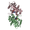
 Assembly
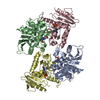


 Type: RNA linking / Mass: 443.201 Da / Num. of mol.: 4 / Source method: obtained synthetically / Formula: C10H15N5O11P2 / Comment: GDP, energy-carrying molecule*YM
 Num. of mol.: 6 / Source method: obtained synthetically
 Mass: 18.015 Da / Num. of mol.: 78 / Source method: isolated from a natural source / Formula: H2O
 Sample preparation
 Processing