 Movie
Movie Controller

[English] 日本語
 Yorodumi
- PDB-2yb0: The Crystal Structure of Leishmania major dUTPase in complex deox... -
+ Open data

- Basic information
| Entry | Database: PDB / ID: 2yb0 | ||||||
|---|---|---|---|---|---|---|---|
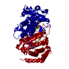
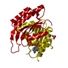
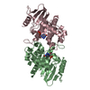
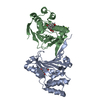
| Title | The Crystal Structure of Leishmania major dUTPase in complex deoxyuridine | ||||||
 Components Components | DUTPASE | ||||||
 Keywords Keywords | HYDROLASE | ||||||
| Function / homology |  Function and homology information Function and homology informationdUTP diphosphatase / dUTP diphosphatase activity / nucleoplasm / metal ion binding Similarity search - Function | ||||||
| Biological species |  LEISHMANIA MAJOR (eukaryote) LEISHMANIA MAJOR (eukaryote) | ||||||
| Method |  X-RAY DIFFRACTION / SYNCHROTRON / MOLECULAR REPLACEMENT / Resolution: 2.28 Å X-RAY DIFFRACTION / SYNCHROTRON / MOLECULAR REPLACEMENT / Resolution: 2.28 Å | ||||||
 Authors Authors | Hemsworth, G.R. / Moroz, O.V. / Fogg, M.J. / Scott, B. / Bosch-Navarrete, C. / Gonzalez-Pacanowska, D. / Wilson, K.S. | ||||||
 Citation Citation | Journal: J.Biol.Chem. / Year: 2011 Title: The Crystal Structure of the Leishmania Major Deoxyuridine Triphosphate Nucleotidohydrolase in Complex with Nucleotide Analogues, Dump, and Deoxyuridine. Authors: Hemsworth, G.R. / Moroz, O.V. / Fogg, M.J. / Scott, B. / Bosch-Navarrete, C. / Gonzalez-Pacanowska, D. / Wilson, K.S. | ||||||
| History |
|
- Structure visualization
| Structure viewer | Molecule: MolmilJmol/JSmol |
|---|
- Downloads & links
-Download
| PDBx/mmCIF format | 2yb0.cif.gz | 417.8 KB | Display | PDBx/mmCIF format |
|---|---|---|---|---|
| PDB format | pdb2yb0.ent.gz | 347.1 KB | Display | PDB format |
| PDBx/mmJSON format | 2yb0.json.gz | Tree view | PDBx/mmJSON format | |
| Others |  Other downloads Other downloads |
-Validation report
| Arichive directory | https://data.pdbj.org/pub/pdb/validation_reports/yb/2yb0ftp://data.pdbj.org/pub/pdb/validation_reports/yb/2yb0 | HTTPS FTP |
|---|
-Related structure data
| Related structure data |  2cicC  2cjeSC  2yayC  2yazC C: citing same article ( S: Starting model for refinement |
|---|---|
| Similar structure data |


-Links
 PDBj
PDBj
- Assembly
| Deposited unit | 
| ||||||||
|---|---|---|---|---|---|---|---|---|---|
| 1 | 
| ||||||||
| 2 | 
| ||||||||
| Unit cell |
|
-Components
| #1: Protein | Mass: 30609.850 Da / Num. of mol.: 4 Source method: isolated from a genetically manipulated source Source: (gene. exp.) LEISHMANIA MAJOR (eukaryote) / Strain: 252 / Plasmid: PET-YSBLLIC3C / Production host:  #2: Chemical | ChemComp-DUR /   Mass: 228.202 Da / Num. of mol.: 4 / Source method: obtained synthetically / Formula: C9H12N2O5 Mass: 228.202 Da / Num. of mol.: 4 / Source method: obtained synthetically / Formula: C9H12N2O5#3: Chemical | ChemComp-SO4 /   Mass: 96.063 Da / Num. of mol.: 9 / Source method: obtained synthetically / Formula: SO4 Mass: 96.063 Da / Num. of mol.: 9 / Source method: obtained synthetically / Formula: SO4#4: Water | ChemComp-HOH / |  Mass: 18.015 Da / Num. of mol.: 97 / Source method: isolated from a natural source / Formula: H2O Mass: 18.015 Da / Num. of mol.: 97 / Source method: isolated from a natural source / Formula: H2O |
|---|
-Experimental details
-Experiment
| Experiment | Method: X-RAY DIFFRACTION / Number of used crystals: 1 |
|---|
- Sample preparation
| Crystal | Density Matthews: 4.29 Å3/Da / Density % sol: 71.32 % / Description: NONE |
|---|---|
| Crystal grow | Details: 0.1 M NACL, 0.1 M BIS-TRIS PH 6.5, 1.5 M (NH4)2SO4 |
-Data collection
| Diffraction | Mean temperature: 100 K |
|---|---|
| Diffraction source | Source: SYNCHROTRON / Site: Diamond  / Beamline: I04 / Wavelength: 0.976 / Beamline: I04 / Wavelength: 0.976 |
| Detector | Type: ADSC CCD / Detector: CCD / Date: Dec 13, 2009 |
| Radiation | Protocol: SINGLE WAVELENGTH / Monochromatic (M) / Laue (L): M / Scattering type: x-ray |
| Radiation wavelength | Wavelength: 0.976 Å / Relative weight: 1 |
| Reflection | Resolution: 2.28→60.8 Å / Num. obs: 98557 / % possible obs: 100 % / Observed criterion σ(I): 1 / Redundancy: 13.5 % / Rmerge(I) obs: 0.11 / Net I/σ(I): 17.4 |
| Reflection shell | Resolution: 2.28→2.4 Å / Redundancy: 10.5 % / Rmerge(I) obs: 0.71 / Mean I/σ(I) obs: 3 / % possible all: 99.8 |
- Processing
| Software |
| ||||||||||||||||||||||||||||||||||||||||||||||||||||||||||||||||||||||||||||||||||||||||||||||||||||||||||||||||||||||||||||||||||||||||||||||||||||||||||||||||||||||||||||||||||||||
|---|---|---|---|---|---|---|---|---|---|---|---|---|---|---|---|---|---|---|---|---|---|---|---|---|---|---|---|---|---|---|---|---|---|---|---|---|---|---|---|---|---|---|---|---|---|---|---|---|---|---|---|---|---|---|---|---|---|---|---|---|---|---|---|---|---|---|---|---|---|---|---|---|---|---|---|---|---|---|---|---|---|---|---|---|---|---|---|---|---|---|---|---|---|---|---|---|---|---|---|---|---|---|---|---|---|---|---|---|---|---|---|---|---|---|---|---|---|---|---|---|---|---|---|---|---|---|---|---|---|---|---|---|---|---|---|---|---|---|---|---|---|---|---|---|---|---|---|---|---|---|---|---|---|---|---|---|---|---|---|---|---|---|---|---|---|---|---|---|---|---|---|---|---|---|---|---|---|---|---|---|---|---|---|
| Refinement | Method to determine structure: MOLECULAR REPLACEMENT Starting model: PDB ENTRY 2CJE Resolution: 2.28→54.59 Å / Cor.coef. Fo:Fc: 0.947 / Cor.coef. Fo:Fc free: 0.936 / SU B: 9.849 / SU ML: 0.114 / Cross valid method: THROUGHOUT / ESU R: 0.18 / ESU R Free: 0.162 / Stereochemistry target values: MAXIMUM LIKELIHOOD / Details: HYDROGENS HAVE BEEN ADDED IN THE RIDING POSITIONS
| ||||||||||||||||||||||||||||||||||||||||||||||||||||||||||||||||||||||||||||||||||||||||||||||||||||||||||||||||||||||||||||||||||||||||||||||||||||||||||||||||||||||||||||||||||||||
| Solvent computation | Ion probe radii: 0.8 Å / Shrinkage radii: 0.8 Å / VDW probe radii: 1.4 Å / Solvent model: MASK | ||||||||||||||||||||||||||||||||||||||||||||||||||||||||||||||||||||||||||||||||||||||||||||||||||||||||||||||||||||||||||||||||||||||||||||||||||||||||||||||||||||||||||||||||||||||
| Displacement parameters | Biso mean: 85.96 Å2
| ||||||||||||||||||||||||||||||||||||||||||||||||||||||||||||||||||||||||||||||||||||||||||||||||||||||||||||||||||||||||||||||||||||||||||||||||||||||||||||||||||||||||||||||||||||||
| Refinement step | Cycle: LAST / Resolution: 2.28→54.59 Å
| ||||||||||||||||||||||||||||||||||||||||||||||||||||||||||||||||||||||||||||||||||||||||||||||||||||||||||||||||||||||||||||||||||||||||||||||||||||||||||||||||||||||||||||||||||||||
| Refine LS restraints |
|