 Movie
Movie Controller


+ Open data

- Basic information
| Entry | Database: PDB / ID: 4jz7 | ||||||
|---|---|---|---|---|---|---|---|
| Title | Carbamate kinase from Giardia lamblia bound to AMP-PNP | ||||||
 Components Components | Carbamate kinase | ||||||
 Keywords Keywords | TRANSFERASE / modified Rossmann fold / ATP carbamate phosphotransferase / ADP / Mg2+ / carbamoyl phosphate | ||||||
| Function / homology |  Function and homology information Function and homology information | ||||||
| Biological species |  Giardia lamblia (eukaryote) Giardia lamblia (eukaryote) | ||||||
| Method |  X-RAY DIFFRACTION / SYNCHROTRON / MOLECULAR REPLACEMENT / Resolution: 2.6 Å X-RAY DIFFRACTION / SYNCHROTRON / MOLECULAR REPLACEMENT / Resolution: 2.6 Å | ||||||
 Authors Authors | Lim, K. / Herzberg, O. | ||||||
 Citation Citation | Journal: Plos One / Year: 2013 Title: Crystal Structures of Carbamate Kinase from Giardia lamblia Bound with Citric Acid and AMP-PNP. Authors: Lim, K. / Kulakova, L. / Galkin, A. / Herzberg, O. #1: Journal: Acta Crystallogr.,Sect.F / Year: 2010Title: X-ray structure and characterization of carbamate kinase from the human parasite Giardia lamblia. Authors: Galkin, A. / Kulakova, L. / Wu, R. / Nash, T.E. / Dunaway-Mariano, D. / Herzberg, O. | ||||||
| History |
|
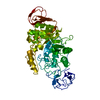
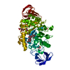
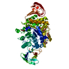
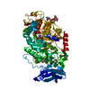
- Structure visualization
| Structure viewer | Molecule: MolmilJmol/JSmol |
|---|
- Downloads & links
-Download
| PDBx/mmCIF format | 4jz7.cif.gz | 244.6 KB | Display | PDBx/mmCIF format |
|---|---|---|---|---|
| PDB format | pdb4jz7.ent.gz | 196.4 KB | Display | PDB format |
| PDBx/mmJSON format | 4jz7.json.gz | Tree view | PDBx/mmJSON format | |
| Others |  Other downloads Other downloads |
-Validation report
| Arichive directory | https://data.pdbj.org/pub/pdb/validation_reports/jz/4jz7ftp://data.pdbj.org/pub/pdb/validation_reports/jz/4jz7 | HTTPS FTP |
|---|
-Related structure data
| Related structure data | 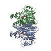 4jz8C 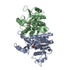 4jz9C 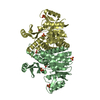 2we4S 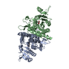 3kzfS S: Starting model for refinement C: citing same article ( |
|---|---|
| Similar structure data |


-Links
 PDBj
PDBj


- Assembly
| Deposited unit | 
| ||||||||
|---|---|---|---|---|---|---|---|---|---|
| 1 | 
| ||||||||
| 2 | 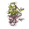
| ||||||||
| Unit cell |
|
-Components
| #1: Protein | Mass: 33975.219 Da / Num. of mol.: 4 Source method: isolated from a genetically manipulated source Source: (gene. exp.) Giardia lamblia (eukaryote) / Strain: ATCC 50803 / WB clone C6 / Gene: GL50803_16453 / Plasmid: pDEST-HisMBP / Production host:  #2: Chemical | ChemComp-ANP /   Mass: 506.196 Da / Num. of mol.: 4 / Source method: obtained synthetically / Formula: C10H17N6O12P3 / Comment: AMP-PNP, energy-carrying molecule analogue*YM Mass: 506.196 Da / Num. of mol.: 4 / Source method: obtained synthetically / Formula: C10H17N6O12P3 / Comment: AMP-PNP, energy-carrying molecule analogue*YM#3: Water | ChemComp-HOH / |  Mass: 18.015 Da / Num. of mol.: 475 / Source method: isolated from a natural source / Formula: H2O Mass: 18.015 Da / Num. of mol.: 475 / Source method: isolated from a natural source / Formula: H2O |
|---|
-Experimental details
-Experiment
| Experiment | Method: X-RAY DIFFRACTION / Number of used crystals: 1 |
|---|
- Sample preparation
| Crystal | Density Matthews: 2.47 Å3/Da / Density % sol: 50.14 % |
|---|---|
| Crystal grow | Temperature: 295 K / Method: vapor diffusion, hanging drop / pH: 5 Details: 0.4 M ammonium citrate dibasic, pH 5.0, 21% PEG 3350, crystal soaked in 50mM AMP-PNP for 4 hours, VAPOR DIFFUSION, HANGING DROP, temperature 295K |
-Data collection
| Diffraction | Mean temperature: 100 K |
|---|---|
| Diffraction source | Source: SYNCHROTRON / Site: APS  / Beamline: 23-ID-B / Wavelength: 1.0332 Å / Beamline: 23-ID-B / Wavelength: 1.0332 Å |
| Detector | Type: MARMOSAIC 300 mm CCD / Detector: CCD / Date: Jun 8, 2011 / Details: mirrors |
| Radiation | Monochromator: Double crystal cryo-cooled Si(111) / Protocol: SINGLE WAVELENGTH / Monochromatic (M) / Laue (L): M / Scattering type: x-ray |
| Radiation wavelength | Wavelength: 1.0332 Å / Relative weight: 1 |
| Reflection | Resolution: 2.6→48.9 Å / Num. all: 40467 / Num. obs: 40467 / % possible obs: 99.2 % / Observed criterion σ(F): 0 / Observed criterion σ(I): 0 / Redundancy: 3.2 % / Biso Wilson estimate: 18 Å2 / Rmerge(I) obs: 0.093 / Net I/σ(I): 8.1 |
| Reflection shell | Resolution: 2.6→2.73 Å / Redundancy: 3.1 % / Rmerge(I) obs: 0.272 / Mean I/σ(I) obs: 3.6 / Num. unique all: 5834 / % possible all: 99.2 |
- Processing
| Software |
| ||||||||||||||||||||
|---|---|---|---|---|---|---|---|---|---|---|---|---|---|---|---|---|---|---|---|---|---|
| Refinement | Method to determine structure: MOLECULAR REPLACEMENT Starting model: PDB ENTRIES 3KZF AND 2WE4 Resolution: 2.6→48.9 Å / Isotropic thermal model: ISOTROPIC / Cross valid method: THROUGHOUT / σ(F): 0 / Stereochemistry target values: Engh & Huber
| ||||||||||||||||||||
| Displacement parameters | Biso mean: 24 Å2 | ||||||||||||||||||||
| Refinement step | Cycle: LAST / Resolution: 2.6→48.9 Å
| ||||||||||||||||||||
| Refine LS restraints |
| ||||||||||||||||||||
| LS refinement shell | Resolution: 2.6→2.73 Å
|
