 Movie
Movie Controller

[English] 日本語
 Yorodumi
- PDB-2grk: Crystal structure of ectromelia virus EVM1 chemokine binding protein -
+ Open data

- Basic information
| Entry | Database: PDB / ID: 2grk | ||||||
|---|---|---|---|---|---|---|---|
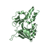
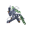
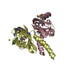
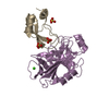
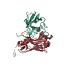
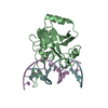
| Title | Crystal structure of ectromelia virus EVM1 chemokine binding protein | ||||||
 Components Components | EVM001 | ||||||
 Keywords Keywords | VIRAL PROTEIN / chemokine binding protein / immune system | ||||||
| Function / homology |  Function and homology information Function and homology informationChemokine-binding protein, viral / Major secreted virus protein / Viral Chemokine Inhibitor; Chain A / Major secreted virus protein, 35kDa / Poxvirus chemokine inhibitor superfamily / Viral chemokine binding protein / Prokaryotic membrane lipoprotein lipid attachment site profile. / Sandwich / Mainly Beta Similarity search - Domain/homology | ||||||
| Biological species |  Ectromelia virus Ectromelia virus | ||||||
| Method |  X-RAY DIFFRACTION / MOLECULAR REPLACEMENT / Resolution: 2.6 Å X-RAY DIFFRACTION / MOLECULAR REPLACEMENT / Resolution: 2.6 Å | ||||||
 Authors Authors | Arnold, P.L. / Fremont, D.H. | ||||||
 Citation Citation | Journal: J.Virol. / Year: 2006 Title: Structural determinants of chemokine binding by an ectromelia virus-encoded decoy receptor. Authors: Arnold, P.L. / Fremont, D.H. | ||||||
| History |
|
- Structure visualization
| Structure viewer | Molecule: MolmilJmol/JSmol |
|---|
- Downloads & links
-Download
| PDBx/mmCIF format | 2grk.cif.gz | 99.2 KB | Display | PDBx/mmCIF format |
|---|---|---|---|---|
| PDB format | pdb2grk.ent.gz | 76.1 KB | Display | PDB format |
| PDBx/mmJSON format | 2grk.json.gz | Tree view | PDBx/mmJSON format | |
| Others |  Other downloads Other downloads |
-Validation report
| Arichive directory | https://data.pdbj.org/pub/pdb/validation_reports/gr/2grkftp://data.pdbj.org/pub/pdb/validation_reports/gr/2grk | HTTPS FTP |
|---|
-Related structure data
| Related structure data |  1cq3S S: Starting model for refinement |
|---|---|
| Similar structure data |


-Links
 PDBj
PDBj


- Assembly
| Deposited unit | 
| ||||||||
|---|---|---|---|---|---|---|---|---|---|
| 1 | 
| ||||||||
| 2 | 
| ||||||||
| Unit cell |
|
-Components
| #1: Protein | Mass: 24684.574 Da / Num. of mol.: 2 Source method: isolated from a genetically manipulated source Source: (gene. exp.) Ectromelia virus / Genus: Orthopoxvirus / Strain: Moscow / Gene: EVM001 / Production host:   Spodoptera frugiperda (fall armyworm) / References: UniProt: Q9JFS0 Spodoptera frugiperda (fall armyworm) / References: UniProt: Q9JFS0#2: Water | ChemComp-HOH / |  Mass: 18.015 Da / Num. of mol.: 122 / Source method: isolated from a natural source / Formula: H2O Mass: 18.015 Da / Num. of mol.: 122 / Source method: isolated from a natural source / Formula: H2OHas protein modification | Y | |
|---|
-Experimental details
-Experiment
| Experiment | Method: X-RAY DIFFRACTION / Number of used crystals: 1 |
|---|
- Sample preparation
| Crystal | Density Matthews: 2.29 Å3/Da / Density % sol: 46.27 % |
|---|---|
| Crystal grow | Temperature: 293 K / pH: 6.2 Details: 2 M ammonium sulfate, 3% ethylene glycol, 0.1 M cacodylate, pH 6.2, VAPOR DIFFUSION, HANGING DROP, temperature 293.0K, pH 6.20 |
-Data collection
| Diffraction | Mean temperature: 100 K |
|---|---|
| Diffraction source | Source: ROTATING ANODE / Type: RIGAKU RU300 / Wavelength: 1.5418 |
| Detector | Type: RIGAKU RAXIS IV / Detector: IMAGE PLATE / Date: May 1, 2003 |
| Radiation | Monochromator: NI FILTER / Protocol: SINGLE WAVELENGTH / Monochromatic (M) / Laue (L): M / Scattering type: x-ray |
| Radiation wavelength | Wavelength: 1.5418 Å / Relative weight: 1 |
| Reflection | Resolution: 2.6→20 Å / Num. obs: 13573 / % possible obs: 97.6 % / Observed criterion σ(I): -3 / Redundancy: 2.9 % / Biso Wilson estimate: 48.2 Å2 / Rmerge(I) obs: 0.112 / Rsym value: 0.085 / Net I/σ(I): 9.547 |
| Reflection shell | Resolution: 2.6→2.72 Å / Rmerge(I) obs: 0.426 / Mean I/σ(I) obs: 2.36 / Rsym value: 0.43 / % possible all: 96.4 |
- Processing
| Software |
| ||||||||||||||||||||||||||||||||||||||||||||||||||||||||||||||||||||||||||||||||
|---|---|---|---|---|---|---|---|---|---|---|---|---|---|---|---|---|---|---|---|---|---|---|---|---|---|---|---|---|---|---|---|---|---|---|---|---|---|---|---|---|---|---|---|---|---|---|---|---|---|---|---|---|---|---|---|---|---|---|---|---|---|---|---|---|---|---|---|---|---|---|---|---|---|---|---|---|---|---|---|---|---|
| Refinement | Method to determine structure: MOLECULAR REPLACEMENT Starting model: PDB ENTRY 1CQ3 Resolution: 2.6→19.88 Å / Rfactor Rfree error: 0.011 / Data cutoff high absF: 148448.609 / Data cutoff low absF: 0 / Isotropic thermal model: RESTRAINED / Cross valid method: THROUGHOUT / σ(F): 0 / Stereochemistry target values: ENGH & HUBER
| ||||||||||||||||||||||||||||||||||||||||||||||||||||||||||||||||||||||||||||||||
| Solvent computation | Solvent model: FLAT MODEL / Bsol: 24.26 Å2 / ksol: 0.33 e/Å3 | ||||||||||||||||||||||||||||||||||||||||||||||||||||||||||||||||||||||||||||||||
| Displacement parameters | Biso mean: 42.2 Å2
| ||||||||||||||||||||||||||||||||||||||||||||||||||||||||||||||||||||||||||||||||
| Refine analyze |
| ||||||||||||||||||||||||||||||||||||||||||||||||||||||||||||||||||||||||||||||||
| Refinement step | Cycle: LAST / Resolution: 2.6→19.88 Å
| ||||||||||||||||||||||||||||||||||||||||||||||||||||||||||||||||||||||||||||||||
| Refine LS restraints |
| ||||||||||||||||||||||||||||||||||||||||||||||||||||||||||||||||||||||||||||||||
| LS refinement shell | Resolution: 2.6→2.76 Å / Rfactor Rfree error: 0.041 / Total num. of bins used: 6
| ||||||||||||||||||||||||||||||||||||||||||||||||||||||||||||||||||||||||||||||||
| Xplor file |
|
