 Movie
Movie Controller

[English] 日本語
 Yorodumi
- PDB-1qnz: NMR structure of the 0.5b anti-HIV antibody complex with the gp12... -
+ Open data

- Basic information
| Entry | Database: PDB / ID: 1qnz | ||||||
|---|---|---|---|---|---|---|---|
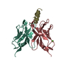
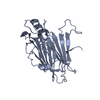
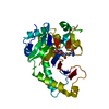
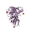
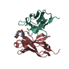
| Title | NMR structure of the 0.5b anti-HIV antibody complex with the gp120 V3 peptide | ||||||
 Components Components |
| ||||||
 Keywords Keywords | IMMUNE SYSTEM / ANTIBODY / V3 PEPTIDE / BINDING SITE | ||||||
| Function / homology |  Function and homology information Function and homology informationmembrane fusion involved in viral entry into host cell / immunoglobulin mediated immune response / immunoglobulin complex / antigen binding / adaptive immune response / immune response / viral envelope / symbiont entry into host cell / virion attachment to host cell / virion membrane ...membrane fusion involved in viral entry into host cell / immunoglobulin mediated immune response / immunoglobulin complex / antigen binding / adaptive immune response / immune response / viral envelope / symbiont entry into host cell / virion attachment to host cell / virion membrane / extracellular region / plasma membrane Similarity search - Function | ||||||
| Biological species |    HUMAN IMMUNODEFICIENCY VIRUS 1 HUMAN IMMUNODEFICIENCY VIRUS 1 | ||||||
| Method | SOLUTION NMR | ||||||
| Model type details | MINIMIZED AVERAGE | ||||||
 Authors Authors | Tugarinov, V. / Zvi, A. / Levy, R. / Hayek, Y. / Matsushita, S. / Anglister, J. | ||||||
 Citation Citation | Journal: Structure / Year: 2000 Title: NMR Structure of an Anti-Gp120 Antibody Complex with a V3 Peptide Reveals a Surface Important for Co-Receptor Binding Authors: Tugarinov, V. / Zvi, A. / Levy, R. / Hayek, Y. / Matsushita, S. / Anglister, J. | ||||||
| History |
|
- Structure visualization
| Structure viewer | Molecule: MolmilJmol/JSmol |
|---|
- Downloads & links
-Download
| PDBx/mmCIF format | 1qnz.cif.gz | 87.9 KB | Display | PDBx/mmCIF format |
|---|---|---|---|---|
| PDB format | pdb1qnz.ent.gz | 67.5 KB | Display | PDB format |
| PDBx/mmJSON format | 1qnz.json.gz | Tree view | PDBx/mmJSON format | |
| Others |  Other downloads Other downloads |
-Validation report
| Arichive directory | https://data.pdbj.org/pub/pdb/validation_reports/qn/1qnzftp://data.pdbj.org/pub/pdb/validation_reports/qn/1qnz | HTTPS FTP |
|---|
-Related structure data
| Similar structure data |
|---|


-Links
 PDBj
PDBj


- Assembly
| Deposited unit | 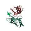
| |||||||||
|---|---|---|---|---|---|---|---|---|---|---|
| 1 |
| |||||||||
| NMR ensembles |
|
-Components
| #1: Antibody | Mass: 13323.770 Da / Num. of mol.: 1 / Fragment: FV Source method: isolated from a genetically manipulated source Source: (gene. exp.)  |
|---|---|
| #2: Antibody | Mass: 12267.469 Da / Num. of mol.: 1 / Fragment: FV Source method: isolated from a genetically manipulated source Source: (gene. exp.) |
| #3: Protein/peptide | Mass: 2017.406 Da / Num. of mol.: 1 / Fragment: V3 PEPTIDE / Source method: obtained synthetically / Source: (synth.) HUMAN IMMUNODEFICIENCY VIRUS 1 / References: UniProt: Q79416 |
| Has protein modification | Y |
-Experimental details
-Experiment
| Experiment | Method: SOLUTION NMR |
|---|---|
| NMR details | Text: THE STRUCTURE OF THE COMPLEX WAS DETERMINED BY ALLOWING THE CDRS OF THE 0.5B ANTIBODY AND THE V3 PEPTIDE RESIDUES TO MOVE WHILE THE FRAMEWORK OF THE ANTIBODY WAS MODELED AS DESCRIBED. |
- Sample preparation
| Sample conditions | Ionic strength: 10 mM sodium phosphate buffer mM / pH: 7.15 / Pressure: 1 atm / Temperature: 305 K |
|---|---|
| Crystal grow | *PLUS Method: other / Details: NMR |
-NMR measurement
| NMR spectrometer |
|
|---|
- Processing
| NMR software |
| ||||||||||||||||||||
|---|---|---|---|---|---|---|---|---|---|---|---|---|---|---|---|---|---|---|---|---|---|
| Refinement | Software ordinal: 1 Details: THE STRUCTURE OF THE COMPLEX WAS DETERMINED ALLOWING THE ANTIBODY CDR RESIDUES AND THE COMPLEXED V3 PEPTIDE RESIDUES TO MOVE, WHILE THE ANTIBODY FRAMEWORK WAS HELD FIXED DURING REFINEMENT ...Details: THE STRUCTURE OF THE COMPLEX WAS DETERMINED ALLOWING THE ANTIBODY CDR RESIDUES AND THE COMPLEXED V3 PEPTIDE RESIDUES TO MOVE, WHILE THE ANTIBODY FRAMEWORK WAS HELD FIXED DURING REFINEMENT AND MODELED AS DESCRIBED PREVIOUSLY | ||||||||||||||||||||
| NMR ensemble | Conformers submitted total number: 1 |
 X-PLOR