 Movie
Movie Controller

[English] 日本語
 Yorodumi
- PDB-3u6u: Crystal structure of the putative acetylglutamate kinase from the... -
+ Open data

- Basic information
| Entry | Database: PDB / ID: 3u6u | |||||||||
|---|---|---|---|---|---|---|---|---|---|---|
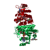
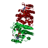
| Title | Crystal structure of the putative acetylglutamate kinase from thermus thermophilus | |||||||||
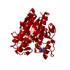
 Components Components | Putative acetylglutamate kinase | |||||||||
 Keywords Keywords | TRANSFERASE / Structural Genomics / RIKEN Structural Genomics/Proteomics Initiative / RSGI / PUTATIVE ACETYLGLUTAMATE KINASE / THERMUS THERMOPHILUS / NPPSFA / NATIONAL PROJECT ON PROTEIN STRUCTURAL AND FUNCTIONAL ANALYSES | |||||||||
| Function / homology |  Function and homology information Function and homology information[amino-group carrier protein]-L-2-aminoadipate 6-kinase / N2-acetyl-L-aminoadipate kinase activity / acetylglutamate kinase activity / : / L-arginine biosynthetic process / ATP binding / cytoplasm Similarity search - Function | |||||||||
| Biological species |   Thermus thermophilus (bacteria) Thermus thermophilus (bacteria) | |||||||||
| Method |  X-RAY DIFFRACTION / SYNCHROTRON / SAD / Resolution: 1.92 Å X-RAY DIFFRACTION / SYNCHROTRON / SAD / Resolution: 1.92 Å | |||||||||
 Authors Authors | Karthe, P. / Kumarevel, T.S. / Kuramitsu, S. / Yokoyama, S. / RIKEN Structural Genomics/Proteomics Initiative (RSGI) | |||||||||
 Citation Citation | Journal: To be Published Title: The structure of first dimeric archaeal N-acetyl glutamate kinase reveals an intermediate conformation of the enzyme in the catalytic cycle Authors: Ramya, S. / Preethi, R. / Kuramitsu, S. / Yokoyama, S. / Kumarevel, T.S. / Karthe, P. | |||||||||
| History |
|
- Structure visualization
| Structure viewer | Molecule: MolmilJmol/JSmol |
|---|
- Downloads & links
-Download
| PDBx/mmCIF format | 3u6u.cif.gz | 113.1 KB | Display | PDBx/mmCIF format |
|---|---|---|---|---|
| PDB format | pdb3u6u.ent.gz | 88 KB | Display | PDB format |
| PDBx/mmJSON format | 3u6u.json.gz | Tree view | PDBx/mmJSON format | |
| Others |  Other downloads Other downloads |
-Validation report
| Arichive directory | https://data.pdbj.org/pub/pdb/validation_reports/u6/3u6uftp://data.pdbj.org/pub/pdb/validation_reports/u6/3u6u | HTTPS FTP |
|---|
-Related structure data
| Similar structure data | |
|---|---|
| Other databases |


-Links
 PDBj
PDBj- Assembly
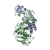
| Deposited unit | 
| ||||||||
|---|---|---|---|---|---|---|---|---|---|
| 1 |
| ||||||||
| Unit cell |
|
-Components
| #1: Protein | Mass: 29092.613 Da / Num. of mol.: 2 Source method: isolated from a genetically manipulated source Source: (gene. exp.) Thermus thermophilus (bacteria) / Strain: HB8 / Gene: TTHA1903 / Plasmid: pET-11a / Production host: #2: Chemical | ChemComp-SO4 /   Mass: 96.063 Da / Num. of mol.: 4 / Source method: obtained synthetically / Formula: SO4 Mass: 96.063 Da / Num. of mol.: 4 / Source method: obtained synthetically / Formula: SO4#3: Water | ChemComp-HOH / |  Mass: 18.015 Da / Num. of mol.: 250 / Source method: isolated from a natural source / Formula: H2O Mass: 18.015 Da / Num. of mol.: 250 / Source method: isolated from a natural source / Formula: H2O |
|---|
-Experimental details
-Experiment
| Experiment | Method: X-RAY DIFFRACTION / Number of used crystals: 1 |
|---|
- Sample preparation
| Crystal | Density Matthews: 4.78 Å3/Da / Density % sol: 74.25 % / Description: the file contains Friedel pairs |
|---|---|
| Crystal grow | Temperature: 293 K / Method: vapor diffusion, sitting drop / pH: 5.6 Details: 0.5M AMMONIUM SULFATE, 0.1M TRI SODIUM CITRATE DEHYDRATE, 1.0M LITHIUM SULFATE MONOHYDRATE, pH 5.6, VAPOR DIFFUSION, SITTING DROP, temperature 293K |
-Data collection
| Diffraction | Mean temperature: 180 K |
|---|---|
| Diffraction source | Source: SYNCHROTRON / Site: SPring-8  / Beamline: BL32B2 / Wavelength: 0.97937 Å / Beamline: BL32B2 / Wavelength: 0.97937 Å |
| Detector | Type: ADSC QUANTUM 210 / Detector: CCD / Date: Dec 25, 2006 |
| Radiation | Monochromator: Si / Protocol: SINGLE WAVELENGTH / Monochromatic (M) / Laue (L): M / Scattering type: x-ray |
| Radiation wavelength | Wavelength: 0.97937 Å / Relative weight: 1 |
| Reflection | Resolution: 1.92→50 Å / Num. all: 163557 / Num. obs: 163557 / % possible obs: 95.4 % / Observed criterion σ(F): 2 / Observed criterion σ(I): 2 / Redundancy: 9.5 % / Biso Wilson estimate: 21.5 Å2 / Rmerge(I) obs: 0.075 |
| Reflection shell | Resolution: 1.92→1.99 Å / Redundancy: 2.2 % / Rmerge(I) obs: 0.601 / Num. unique all: 4894 / % possible all: 58.2 |
- Processing
| Software |
| ||||||||||||||||||||||||||||||||||||
|---|---|---|---|---|---|---|---|---|---|---|---|---|---|---|---|---|---|---|---|---|---|---|---|---|---|---|---|---|---|---|---|---|---|---|---|---|---|
| Refinement | Method to determine structure: SAD / Resolution: 1.92→19.9 Å / Rfactor Rfree error: 0.003 / Data cutoff high absF: 466950.16 / Data cutoff low absF: 0 / Isotropic thermal model: RESTRAINED / Cross valid method: THROUGHOUT / σ(F): 2 / Stereochemistry target values: Engh & Huber / Details: The file contains Friedel pairs
| ||||||||||||||||||||||||||||||||||||
| Solvent computation | Solvent model: FLAT MODEL / Bsol: 53.0817 Å2 / ksol: 0.380042 e/Å3 | ||||||||||||||||||||||||||||||||||||
| Displacement parameters | Biso mean: 37.9 Å2
| ||||||||||||||||||||||||||||||||||||
| Refine analyze |
| ||||||||||||||||||||||||||||||||||||
| Refinement step | Cycle: LAST / Resolution: 1.92→19.9 Å
| ||||||||||||||||||||||||||||||||||||
| Refine LS restraints |
| ||||||||||||||||||||||||||||||||||||
| LS refinement shell | Resolution: 1.92→2.04 Å / Rfactor Rfree error: 0.011 / Total num. of bins used: 6
| ||||||||||||||||||||||||||||||||||||
| Xplor file |
|
