Mass: 616.487 Da / Num. of mol.: 1 / Source method: obtained synthetically / Formula: C34H32FeN4O4
Compound details
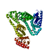
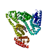
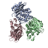
SERUM ALBUMIN, REGULATES THE COLLOIDAL OSMOTIC PRESSURE OF IT BINDS TO WATER, CA++, NA+, K+, FATTY ...SERUM ALBUMIN, REGULATES THE COLLOIDAL OSMOTIC PRESSURE OF IT BINDS TO WATER, CA++, NA+, K+, FATTY ACIDS, HORMONES, BILIRUBIN AND DRUGS
Has protein modification
Y
-
Experimental details
-
Experiment
Experiment
Method: X-RAY DIFFRACTION / Number of used crystals: 1
-
Sample preparation
Crystal
Density Matthews: 2.66 Å3/Da / Density % sol: 54 %
Crystal grow
pH: 7 / Details: pH 7.00
Crystal grow
*PLUS
Temperature: 4 ℃ / Method: vapor diffusion, sitting drop
Resolution: 3.2→27.23 Å / Rfactor Rfree error: 0.012 / Data cutoff high absF: 1526339.7 / Data cutoff low absF: 0 / Isotropic thermal model: GROUP / Cross valid method: THROUGHOUT / σ(F): 0 Details: BULK SOLVENT MODEL USED RESIDUES ASP 1, ALA 2 AND LEU 585 ARE DISORDERED AND HAVE NOT BEEN INCLUDED IN THE MODEL.
In the structure databanks used in Yorodumi, some data are registered as the other names, "COVID-19 virus" and "2019-nCoV". Here are the details of the virus and the list of structure data.
Jan 31, 2019. EMDB accession codes are about to change! (news from PDBe EMDB page)
EMDB accession codes are about to change! (news from PDBe EMDB page)
The allocation of 4 digits for EMDB accession codes will soon come to an end. Whilst these codes will remain in use, new EMDB accession codes will include an additional digit and will expand incrementally as the available range of codes is exhausted. The current 4-digit format prefixed with “EMD-” (i.e. EMD-XXXX) will advance to a 5-digit format (i.e. EMD-XXXXX), and so on. It is currently estimated that the 4-digit codes will be depleted around Spring 2019, at which point the 5-digit format will come into force.
The EM Navigator/Yorodumi systems omit the EMD- prefix.
Related info.:Q: What is EMD? / ID/Accession-code notation in Yorodumi/EM Navigator
Yorodumi is a browser for structure data from EMDB, PDB, SASBDB, etc.
This page is also the successor to EM Navigator detail page, and also detail information page/front-end page for Omokage search.
The word "yorodu" (or yorozu) is an old Japanese word meaning "ten thousand". "mi" (miru) is to see.
Related info.:EMDB / PDB / SASBDB / Comparison of 3 databanks / Yorodumi Search / Aug 31, 2016. New EM Navigator & Yorodumi / Yorodumi Papers / Jmol/JSmol / Function and homology information / Changes in new EM Navigator and Yorodumi
 Movie
Movie Controller

 Yorodumi
 Open data
 Basic information
 Components
 Keywords
 Function and homology information
 HOMO SAPIENS (human)
 X-RAY DIFFRACTION /
  Authors
 Citation
 Structure visualization
 Downloads & links
 Other downloads


 PDBj
PDBj


 Assembly
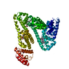
 PICHIA PASTORIS (fungus) / References: UniProt: P02768
 Mass: 228.371 Da / Num. of mol.: 6 / Source method: obtained synthetically / Formula: C14H28O2
 Mass: 616.487 Da / Num. of mol.: 1 / Source method: obtained synthetically / Formula: C34H32FeN4O4
 Sample preparation
 / Beamline: PX9.6 / Wavelength: 0.87
  Processing