 Movie
Movie Controller

[English] 日本語
 Yorodumi
- PDB-1e7b: Crystal structure of human serum albumin complexed with the gener... -
+ Open data

- Basic information
| Entry | Database: PDB / ID: 1e7b | ||||||
|---|---|---|---|---|---|---|---|
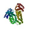
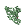
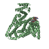
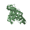
| Title | Crystal structure of human serum albumin complexed with the general anesthetic halothane | ||||||
 Components Components | SERUM ALBUMIN | ||||||
 Keywords Keywords | CARRIER PROTEIN / ALBUMIN / GENERAL ANESTHETIC / HALOTHANE | ||||||
| Function / homology |  Function and homology information Function and homology informationbilirubin transport / Ciprofloxacin ADME / exogenous protein binding / cellular response to calcium ion starvation / enterobactin binding / Heme biosynthesis / HDL remodeling / molecular carrier activity / negative regulation of mitochondrial depolarization / Prednisone ADME ...bilirubin transport / Ciprofloxacin ADME / exogenous protein binding / cellular response to calcium ion starvation / enterobactin binding / Heme biosynthesis / HDL remodeling / molecular carrier activity / negative regulation of mitochondrial depolarization / Prednisone ADME / Heme degradation / Aspirin ADME / antioxidant activity / Scavenging of heme from plasma / toxic substance binding / Recycling of bile acids and salts / platelet alpha granule lumen / fatty acid binding / cellular response to starvation / Post-translational protein phosphorylation / Cytoprotection by HMOX1 / response to nutrient levels / Regulation of Insulin-like Growth Factor (IGF) transport and uptake by Insulin-like Growth Factor Binding Proteins (IGFBPs) / Platelet degranulation / pyridoxal phosphate binding / protein-folding chaperone binding / blood microparticle / endoplasmic reticulum lumen / copper ion binding / Golgi apparatus / endoplasmic reticulum / protein-containing complex / : / DNA binding / extracellular exosome / extracellular region / identical protein binding / nucleus Similarity search - Function | ||||||
| Biological species |  HOMO SAPIENS (human) HOMO SAPIENS (human) | ||||||
| Method |  X-RAY DIFFRACTION / SYNCHROTRON / MOLECULAR REPLACEMENT / Resolution: 2.38 Å X-RAY DIFFRACTION / SYNCHROTRON / MOLECULAR REPLACEMENT / Resolution: 2.38 Å | ||||||
 Authors Authors | Bhattacharya, A.A. / Curry, S. / Franks, N.P. | ||||||
 Citation Citation | Journal: J.Biol.Chem. / Year: 2000 Title: Binding of the General Anesthetics Propofol and Halothane to Human Serum Albumin. High Resolution Crystal Structures Authors: Bhattacharya, A.A. / Curry, S. / Franks, N.P. | ||||||
| History |
|
- Structure visualization
| Structure viewer | Molecule: MolmilJmol/JSmol |
|---|
- Downloads & links
-Download
| PDBx/mmCIF format | 1e7b.cif.gz | 227.1 KB | Display | PDBx/mmCIF format |
|---|---|---|---|---|
| PDB format | pdb1e7b.ent.gz | 179.4 KB | Display | PDB format |
| PDBx/mmJSON format | 1e7b.json.gz | Tree view | PDBx/mmJSON format | |
| Others |  Other downloads Other downloads |
-Validation report
| Arichive directory | https://data.pdbj.org/pub/pdb/validation_reports/e7/1e7bftp://data.pdbj.org/pub/pdb/validation_reports/e7/1e7b | HTTPS FTP |
|---|
-Related structure data
| Related structure data |  1e78C  1e7aC  1e7cC  1ao6S S: Starting model for refinement C: citing same article ( |
|---|---|
| Similar structure data |


-Links
 PDBj
PDBj


- Assembly
| Deposited unit | 
| ||||||||
|---|---|---|---|---|---|---|---|---|---|
| 1 | 
| ||||||||
| 2 | 
| ||||||||
| Unit cell |
| ||||||||
| Noncrystallographic symmetry (NCS) | NCS oper: (Code: given Matrix: (0.4415, 0.8968, 0.0291), Vector: |
-Components
| #1: Protein | Mass: 66571.219 Da / Num. of mol.: 2 Source method: isolated from a genetically manipulated source Source: (gene. exp.) HOMO SAPIENS (human) / Production host:  #2: Chemical | ChemComp-HLT /   Mass: 197.382 Da / Num. of mol.: 6 / Source method: obtained synthetically / Formula: C2HBrClF3 Mass: 197.382 Da / Num. of mol.: 6 / Source method: obtained synthetically / Formula: C2HBrClF3#3: Water | ChemComp-HOH / |  Mass: 18.015 Da / Num. of mol.: 57 / Source method: isolated from a natural source / Formula: H2O Mass: 18.015 Da / Num. of mol.: 57 / Source method: isolated from a natural source / Formula: H2OCompound details | SERUM ALBUMIN, REGULATES THE COLLOIDAL OSMOTIC PRESSURE OF BLOOD IT BINDS TO WATER, CA++, NA+, K+, ...SERUM ALBUMIN, REGULATES THE COLLOIDAL OSMOTIC PRESSURE OF BLOOD IT BINDS TO WATER, CA++, NA+, K+, FATTY ACIDS, HORMONES, BILIRUBIN AND DRUGS | Has protein modification | Y | Sequence details | RESIDUES 1-24 IN P02768 ENTRY ARE SIGNAL SEQUENCE. RESIDUE 1 IN STRUCTURE COORDINATES IS EQUIVALENT ...RESIDUES 1-24 IN P02768 ENTRY ARE SIGNAL SEQUENCE. RESIDUE 1 IN STRUCTURE COORDINATE | |
|---|
-Experimental details
-Experiment
| Experiment | Method: X-RAY DIFFRACTION / Number of used crystals: 1 |
|---|
- Sample preparation
| Crystal | Density Matthews: 2.42 Å3/Da / Density % sol: 49.27 % | |||||||||||||||
|---|---|---|---|---|---|---|---|---|---|---|---|---|---|---|---|---|
| Crystal grow | pH: 7 / Details: pH 7.00 | |||||||||||||||
| Crystal grow | *PLUS Temperature: 4 ℃ / Method: vapor diffusion, sitting drop | |||||||||||||||
| Components of the solutions | *PLUS
|
-Data collection
| Diffraction | Mean temperature: 298 K |
|---|---|
| Diffraction source | Source: SYNCHROTRON / Site: EMBL/DESY, HAMBURG  / Beamline: X11 / Wavelength: 0.909 / Beamline: X11 / Wavelength: 0.909 |
| Detector | Type: MARRESEARCH / Detector: IMAGE PLATE / Date: Jul 15, 1999 / Details: MIRRORS |
| Radiation | Monochromator: GE(111) / Protocol: SINGLE WAVELENGTH / Monochromatic (M) / Laue (L): M / Scattering type: x-ray |
| Radiation wavelength | Wavelength: 0.909 Å / Relative weight: 1 |
| Reflection | Resolution: 2.38→14.97 Å / Num. obs: 48001 / % possible obs: 95.7 % / Observed criterion σ(I): 0 / Redundancy: 1.9 % / Biso Wilson estimate: 39.3 Å2 / Rmerge(I) obs: 0.049 / Rsym value: 0.049 / Net I/σ(I): 8.1 |
| Reflection shell | Resolution: 2.38→2.51 Å / Redundancy: 1.8 % / Rmerge(I) obs: 0.267 / Mean I/σ(I) obs: 2.2 / Rsym value: 0.267 / % possible all: 87.7 |
| Reflection shell | *PLUS % possible obs: 87.7 % |
- Processing
| Software |
| ||||||||||||||||||||||||||||||||||||||||||||||||||||||||||||||||||||||||||||||||
|---|---|---|---|---|---|---|---|---|---|---|---|---|---|---|---|---|---|---|---|---|---|---|---|---|---|---|---|---|---|---|---|---|---|---|---|---|---|---|---|---|---|---|---|---|---|---|---|---|---|---|---|---|---|---|---|---|---|---|---|---|---|---|---|---|---|---|---|---|---|---|---|---|---|---|---|---|---|---|---|---|---|
| Refinement | Method to determine structure: MOLECULAR REPLACEMENT Starting model: 1AO6 Resolution: 2.38→17 Å / Rfactor Rfree error: 0.006 / Data cutoff high absF: 10000000 / Data cutoff low absF: 0.001 / Isotropic thermal model: RESTRAINED / Cross valid method: THROUGHOUT / σ(F): 0 Details: BULK SOLVENT MODEL USED REFINEMENT WAS PERFORMED WITH MAX ALLOWABLE TEMPERATURE FACTOR OF 150
| ||||||||||||||||||||||||||||||||||||||||||||||||||||||||||||||||||||||||||||||||
| Displacement parameters | Biso mean: 76.1 Å2
| ||||||||||||||||||||||||||||||||||||||||||||||||||||||||||||||||||||||||||||||||
| Refine analyze |
| ||||||||||||||||||||||||||||||||||||||||||||||||||||||||||||||||||||||||||||||||
| Refinement step | Cycle: LAST / Resolution: 2.38→17 Å
| ||||||||||||||||||||||||||||||||||||||||||||||||||||||||||||||||||||||||||||||||
| Refine LS restraints |
| ||||||||||||||||||||||||||||||||||||||||||||||||||||||||||||||||||||||||||||||||
| LS refinement shell | Resolution: 2.38→2.49 Å / Rfactor Rfree error: 0.026 / Total num. of bins used: 8
| ||||||||||||||||||||||||||||||||||||||||||||||||||||||||||||||||||||||||||||||||
| Xplor file |
| ||||||||||||||||||||||||||||||||||||||||||||||||||||||||||||||||||||||||||||||||
| Software | *PLUS Name: X-PLOR / Version: 3.851 / Classification: refinement | ||||||||||||||||||||||||||||||||||||||||||||||||||||||||||||||||||||||||||||||||
| Refinement | *PLUS Rfactor obs: 0.27 / Rfactor Rwork: 0.27 | ||||||||||||||||||||||||||||||||||||||||||||||||||||||||||||||||||||||||||||||||
| Solvent computation | *PLUS | ||||||||||||||||||||||||||||||||||||||||||||||||||||||||||||||||||||||||||||||||
| Displacement parameters | *PLUS | ||||||||||||||||||||||||||||||||||||||||||||||||||||||||||||||||||||||||||||||||
| Refine LS restraints | *PLUS
|
