- PDB-4emx: Crystal structure analysis of Human Serum Albumin in complex with... -
+
Open data
ID or keywords:
Loading...
-
Basic information
Entry
Database: PDB / ID: 4emx
Title
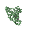
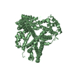
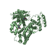
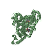
Crystal structure analysis of Human Serum Albumin in complex with chloride anions at cryogenic temperature
Components
Serum albumin
Keywords
PROTEIN BINDING / plasma / drug binding / antioxidant / allostery / redox biology / all alpha / disulfide bond stabilized / human plasma osmotic regulation / transport / extracellular space / free radicals
Function / homology
Function and homology information
bilirubin transport / Ciprofloxacin ADME / exogenous protein binding / cellular response to calcium ion starvation / enterobactin binding / Heme biosynthesis / HDL remodeling / molecular carrier activity / negative regulation of mitochondrial depolarization / Prednisone ADME ...bilirubin transport / Ciprofloxacin ADME / exogenous protein binding / cellular response to calcium ion starvation / enterobactin binding / Heme biosynthesis / HDL remodeling / molecular carrier activity / negative regulation of mitochondrial depolarization / Prednisone ADME / Heme degradation / Aspirin ADME / antioxidant activity / Scavenging of heme from plasma / toxic substance binding / Recycling of bile acids and salts / platelet alpha granule lumen / fatty acid binding / cellular response to starvation / Post-translational protein phosphorylation / Cytoprotection by HMOX1 / response to nutrient levels / Regulation of Insulin-like Growth Factor (IGF) transport and uptake by Insulin-like Growth Factor Binding Proteins (IGFBPs) / Platelet degranulation / pyridoxal phosphate binding / protein-folding chaperone binding / blood microparticle / endoplasmic reticulum lumen / copper ion binding / Golgi apparatus / endoplasmic reticulum / protein-containing complex / : / DNA binding / extracellular exosome / extracellular region / identical protein binding / nucleus Similarity search - Function
Serum Albumin; Chain A, Domain 1 - #10 / Serum albumin/Alpha-fetoprotein/Afamin / ALB/AFP/VDB / Serum albumin, N-terminal / Serum albumin, conserved site / Serum albumin-like / Serum albumin family / Albumin domain signature. / Albumin domain profile. / serum albumin ...Serum Albumin; Chain A, Domain 1 - #10 / Serum albumin/Alpha-fetoprotein/Afamin / ALB/AFP/VDB / Serum albumin, N-terminal / Serum albumin, conserved site / Serum albumin-like / Serum albumin family / Albumin domain signature. / Albumin domain profile. / serum albumin / Serum Albumin; Chain A, Domain 1 / Orthogonal Bundle / Mainly Alpha Similarity search - Domain/homology
Mass: 18.015 Da / Num. of mol.: 228 / Source method: isolated from a natural source / Formula: H2O
Has protein modification
Y
-
Experimental details
-
Experiment
Experiment
Method: X-RAY DIFFRACTION / Number of used crystals: 1
-
Sample preparation
Crystal
Density Matthews: 2.33 Å3/Da / Density % sol: 47.17 %
Crystal grow
Temperature: 293 K / Method: vapor diffusion, hanging drop / pH: 7.5 Details: 2 microL 100 mg/mL protein in 50 mM disodium/monosodium phosphate buffer and 100 mM NaCl were mixed with 2 uL 25% PEG 3350, 50 mM disodium/monosodium phosphate buffer, 100 mM NaCl, pH 7.5, ...Details: 2 microL 100 mg/mL protein in 50 mM disodium/monosodium phosphate buffer and 100 mM NaCl were mixed with 2 uL 25% PEG 3350, 50 mM disodium/monosodium phosphate buffer, 100 mM NaCl, pH 7.5, vapor diffusion, hanging drop, temperature 293K
Protocol: SINGLE WAVELENGTH / Monochromatic (M) / Laue (L): M / Scattering type: x-ray
Radiation wavelength
Wavelength: 1.0809 Å / Relative weight: 1
Reflection
Resolution: 2.28→25.566 Å / Num. all: 50973 / Num. obs: 50973 / % possible obs: 93.1 % / Redundancy: 2.6 % / Rsym value: 0.066 / Net I/σ(I): 9.7
Reflection shell
Diffraction-ID: 1
Resolution (Å)
Redundancy (%)
Rmerge(I) obs
Mean I/σ(I) obs
Num. measured all
Num. unique all
Rsym value
% possible all
2.28-2.4
2.6
0.367
1.9
19776
7579
0.367
95
2.4-2.55
2.6
0.234
2.9
18753
7210
0.234
95
2.55-2.73
2.6
0.163
1.2
17578
6732
0.163
94.7
2.73-2.94
2.6
0.107
6.4
16448
6308
0.107
95
2.94-3.22
2.6
0.078
8.3
14954
5722
0.078
94.3
3.22-3.61
2.6
0.062
9.5
13435
5158
0.062
93.9
3.61-4.16
2.6
0.056
9.3
11823
4512
0.056
92.3
4.16-5.1
2.6
0.05
11.7
9294
3624
0.05
88.9
5.1-7.21
2.7
0.054
11.1
7920
2883
0.054
90.6
7.21-25.723
2.7
0.041
14.8
3401
1245
0.041
72
-
Phasing
Phasing
Method: molecular replacement
-
Processing
Software
Name
Version
Classification
NB
MOSFLM
datareduction
SCALA
3.3.9
datascaling
AMoRE
phasing
REFMAC
refinement
PDB_EXTRACT
3.11
dataextraction
HKL-2000
datacollection
Refinement
Method to determine structure: MOLECULAR REPLACEMENT / Resolution: 2.3→25.5 Å / Cor.coef. Fo:Fc: 0.957 / Cor.coef. Fo:Fc free: 0.927 / WRfactor Rfree: 0.2593 / WRfactor Rwork: 0.2025 / Occupancy max: 1 / Occupancy min: 0.3 / FOM work R set: 0.8281 / SU B: 17.279 / SU ML: 0.204 / SU R Cruickshank DPI: 0.4073 / SU Rfree: 0.2479 / Cross valid method: THROUGHOUT / σ(F): 0 / ESU R: 0.407 / ESU R Free: 0.248 / Stereochemistry target values: MAXIMUM LIKELIHOOD Details: HYDROGENS HAVE BEEN USED IF PRESENT IN THE INPUT U VALUES : WITH TLS ADDED
Rfactor
Num. reflection
% reflection
Selection details
Rfree
0.2381
1010
2 %
RANDOM
Rwork
0.1873
-
-
-
obs
0.1883
49673
93.03 %
-
Solvent computation
Ion probe radii: 0.8 Å / Shrinkage radii: 0.8 Å / VDW probe radii: 1.2 Å / Solvent model: BABINET MODEL WITH MASK
In the structure databanks used in Yorodumi, some data are registered as the other names, "COVID-19 virus" and "2019-nCoV". Here are the details of the virus and the list of structure data.
Jan 31, 2019. EMDB accession codes are about to change! (news from PDBe EMDB page)
EMDB accession codes are about to change! (news from PDBe EMDB page)
The allocation of 4 digits for EMDB accession codes will soon come to an end. Whilst these codes will remain in use, new EMDB accession codes will include an additional digit and will expand incrementally as the available range of codes is exhausted. The current 4-digit format prefixed with “EMD-” (i.e. EMD-XXXX) will advance to a 5-digit format (i.e. EMD-XXXXX), and so on. It is currently estimated that the 4-digit codes will be depleted around Spring 2019, at which point the 5-digit format will come into force.
The EM Navigator/Yorodumi systems omit the EMD- prefix.
Related info.:Q: What is EMD? / ID/Accession-code notation in Yorodumi/EM Navigator
Yorodumi is a browser for structure data from EMDB, PDB, SASBDB, etc.
This page is also the successor to EM Navigator detail page, and also detail information page/front-end page for Omokage search.
The word "yorodu" (or yorozu) is an old Japanese word meaning "ten thousand". "mi" (miru) is to see.
Related info.:EMDB / PDB / SASBDB / Comparison of 3 databanks / Yorodumi Search / Aug 31, 2016. New EM Navigator & Yorodumi / Yorodumi Papers / Jmol/JSmol / Function and homology information / Changes in new EM Navigator and Yorodumi
 Movie
Movie Controller

 Yorodumi
 Open data
 Basic information
 Components
 Keywords
 Function and homology information
 Homo sapiens (human)
 X-RAY DIFFRACTION /
  Authors
 Citation
 Structure visualization
 Downloads & links
 Other downloads


 PDBj
PDBj


 Assembly

 Mass: 35.453 Da / Num. of mol.: 6 / Source method: obtained synthetically / Formula: Cl
 Mass: 18.015 Da / Num. of mol.: 228 / Source method: isolated from a natural source / Formula: H2O
 Sample preparation
 / Beamline: X26C / Wavelength: 1.0809 Å
 Processing