 Movie
Movie Controller


+ Open data

- Basic information
| Entry | Database: PDB / ID: 1bm0 | ||||||
|---|---|---|---|---|---|---|---|
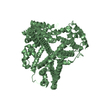
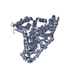
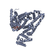
| Title | CRYSTAL STRUCTURE OF HUMAN SERUM ALBUMIN | ||||||
 Components Components | SERUM ALBUMIN | ||||||
 Keywords Keywords | CARRIER PROTEIN / ALBUMIN | ||||||
| Function / homology |  Function and homology information Function and homology informationbilirubin transport / Ciprofloxacin ADME / exogenous protein binding / cellular response to calcium ion starvation / enterobactin binding / Heme biosynthesis / HDL remodeling / molecular carrier activity / negative regulation of mitochondrial depolarization / Prednisone ADME ...bilirubin transport / Ciprofloxacin ADME / exogenous protein binding / cellular response to calcium ion starvation / enterobactin binding / Heme biosynthesis / HDL remodeling / molecular carrier activity / negative regulation of mitochondrial depolarization / Prednisone ADME / Heme degradation / Aspirin ADME / antioxidant activity / Scavenging of heme from plasma / toxic substance binding / Recycling of bile acids and salts / platelet alpha granule lumen / fatty acid binding / cellular response to starvation / Post-translational protein phosphorylation / Cytoprotection by HMOX1 / response to nutrient levels / Regulation of Insulin-like Growth Factor (IGF) transport and uptake by Insulin-like Growth Factor Binding Proteins (IGFBPs) / Platelet degranulation / pyridoxal phosphate binding / protein-folding chaperone binding / blood microparticle / endoplasmic reticulum lumen / copper ion binding / Golgi apparatus / endoplasmic reticulum / protein-containing complex / : / DNA binding / extracellular exosome / extracellular region / identical protein binding / nucleus Similarity search - Function | ||||||
| Biological species |  Homo sapiens (human) Homo sapiens (human) | ||||||
| Method |  X-RAY DIFFRACTION / MOLECULAR REPLACEMENT / Resolution: 2.5 Å X-RAY DIFFRACTION / MOLECULAR REPLACEMENT / Resolution: 2.5 Å | ||||||
 Authors Authors | Sugio, S. / Kashima, A. / Mochizuki, S. / Noda, M. / Kobayashi, K. | ||||||
 Citation Citation | Journal: Protein Eng. / Year: 1999 Title: Crystal structure of human serum albumin at 2.5 A resolution. Authors: Sugio, S. / Kashima, A. / Mochizuki, S. / Noda, M. / Kobayashi, K. #1: Journal: Adv.Protein Chem. / Year: 1994Title: Structure of Serum Albumin Authors: Carter, D.C. / Ho, J.X. #2: Journal: Eur.J.Biochem. / Year: 1994Title: Preliminary Crystallographic Studies of Four Crystal Forms of Serum Albumin Authors: Carter, D.C. / Chang, B. / Ho, J.X. / Keeling, K. / Krishnasami, Z. #3: Journal: Nature / Year: 1993Title: Erratum. Atomic Structure and Chemistry of Human Serum Albumin Authors: He, X.M. / Carter, D.C. #4: Journal: Nature / Year: 1992Title: Atomic Structure and Chemistry of Human Serum Albumin Authors: He, X.M. / Carter, D.C. #5: Journal: Science / Year: 1990Title: Structure of Human Serum Albumin Authors: Carter, D.C. / He, X.M. #6: Journal: Science / Year: 1989Title: Three-Dimensional Structure of Human Serum Albumin Authors: Carter, D.C. / He, X.M. / Munson, S.H. / Twigg, P.D. / Gernert, K.M. / Broom, M.B. / Miller, T.Y. | ||||||
| History |
|
- Structure visualization
| Structure viewer | Molecule: MolmilJmol/JSmol |
|---|
- Downloads & links
-Download
| PDBx/mmCIF format | 1bm0.cif.gz | 230.6 KB | Display | PDBx/mmCIF format |
|---|---|---|---|---|
| PDB format | pdb1bm0.ent.gz | 187.4 KB | Display | PDB format |
| PDBx/mmJSON format | 1bm0.json.gz | Tree view | PDBx/mmJSON format | |
| Others |  Other downloads Other downloads |
-Validation report
| Arichive directory | https://data.pdbj.org/pub/pdb/validation_reports/bm/1bm0ftp://data.pdbj.org/pub/pdb/validation_reports/bm/1bm0 | HTTPS FTP |
|---|
-Related structure data
| Related structure data |  1ao6SC S: Starting model for refinement C: citing same article ( |
|---|---|
| Similar structure data |


-Links
 PDBj
PDBj


- Assembly
| Deposited unit | 
| ||||||||
|---|---|---|---|---|---|---|---|---|---|
| 1 | 
| ||||||||
| 2 | 
| ||||||||
| Unit cell |
|
-Components
| #1: Protein | Mass: 66571.219 Da / Num. of mol.: 2 Source method: isolated from a genetically manipulated source Source: (gene. exp.) Homo sapiens (human) / Tissue: PLASMA / Production host:  Pichia pastoris (fungus) / References: UniProt: P02768 Pichia pastoris (fungus) / References: UniProt: P02768#2: Water | ChemComp-HOH / |  Mass: 18.015 Da / Num. of mol.: 4 / Source method: isolated from a natural source / Formula: H2O Mass: 18.015 Da / Num. of mol.: 4 / Source method: isolated from a natural source / Formula: H2OHas protein modification | Y | |
|---|
-Experimental details
-Experiment
| Experiment | Method: X-RAY DIFFRACTION / Number of used crystals: 5 |
|---|
- Sample preparation
| Crystal | Density Matthews: 2.4 Å3/Da / Density % sol: 48.76 % | |||||||||||||||||||||||||
|---|---|---|---|---|---|---|---|---|---|---|---|---|---|---|---|---|---|---|---|---|---|---|---|---|---|---|
| Crystal grow | pH: 7.5 / Details: pH 7.5 | |||||||||||||||||||||||||
| Crystal | *PLUS Density % sol: 77 % | |||||||||||||||||||||||||
| Crystal grow | *PLUS Temperature: 293 K / Method: vapor diffusion, hanging drop / PH range low: 5.5 / PH range high: 5 | |||||||||||||||||||||||||
| Components of the solutions | *PLUS
|
-Data collection
| Diffraction | Mean temperature: 293 K |
|---|---|
| Diffraction source | Source: ROTATING ANODE / Type: RIGAKU RUH2R / Wavelength: 1.5418 |
| Detector | Type: RIGAKU RAXIS IIC / Detector: IMAGE PLATE / Date: May 14, 1996 / Details: YALE/MSC MIRRORS |
| Radiation | Monochromator: NI FILTER / Monochromatic (M) / Laue (L): M / Scattering type: x-ray |
| Radiation wavelength | Wavelength: 1.5418 Å / Relative weight: 1 |
| Reflection | Resolution: 2.37→93.47 Å / Num. obs: 36149 / % possible obs: 71.3 % / Observed criterion σ(I): 1 / Redundancy: 3.7 % / Biso Wilson estimate: 24.4 Å2 / Rmerge(I) obs: 0.134 / Net I/σ(I): 4.37 |
| Reflection shell | Resolution: 2.37→2.5 Å / Rmerge(I) obs: 0.428 / Mean I/σ(I) obs: 1.52 / % possible all: 36.1 |
| Reflection shell | *PLUS % possible obs: 36 % |
- Processing
| Software |
| ||||||||||||||||||||||||||||||||||||||||||||||||||||||||||||||||||||||||||||||||
|---|---|---|---|---|---|---|---|---|---|---|---|---|---|---|---|---|---|---|---|---|---|---|---|---|---|---|---|---|---|---|---|---|---|---|---|---|---|---|---|---|---|---|---|---|---|---|---|---|---|---|---|---|---|---|---|---|---|---|---|---|---|---|---|---|---|---|---|---|---|---|---|---|---|---|---|---|---|---|---|---|---|
| Refinement | Method to determine structure: MOLECULAR REPLACEMENT Starting model: HUMAN SERUM ALBUMIN (1AO6) Resolution: 2.5→50 Å / Rfactor Rfree error: 0.005 / Data cutoff high absF: 100000 / Data cutoff low absF: 0.1 / Isotropic thermal model: RESTRAINED / Cross valid method: POSTERIORI / σ(F): 0 Details: THE AUTHORS USED BULK SOLVENT CORRECTION AND WEIGHTING SCHEME DEPENDENT ON RESOLUTIONS.
| ||||||||||||||||||||||||||||||||||||||||||||||||||||||||||||||||||||||||||||||||
| Displacement parameters | Biso mean: 40.7 Å2 | ||||||||||||||||||||||||||||||||||||||||||||||||||||||||||||||||||||||||||||||||
| Refine analyze | Luzzati coordinate error obs: 0.3 Å / Luzzati d res low obs: 5 Å | ||||||||||||||||||||||||||||||||||||||||||||||||||||||||||||||||||||||||||||||||
| Refinement step | Cycle: LAST / Resolution: 2.5→50 Å
| ||||||||||||||||||||||||||||||||||||||||||||||||||||||||||||||||||||||||||||||||
| Refine LS restraints |
| ||||||||||||||||||||||||||||||||||||||||||||||||||||||||||||||||||||||||||||||||
| LS refinement shell | Resolution: 2.5→2.59 Å / Rfactor Rfree error: 0.028 / Total num. of bins used: 10
| ||||||||||||||||||||||||||||||||||||||||||||||||||||||||||||||||||||||||||||||||
| Xplor file |
| ||||||||||||||||||||||||||||||||||||||||||||||||||||||||||||||||||||||||||||||||
| Software | *PLUS Name: X-PLOR / Version: 3.1 / Classification: refinement | ||||||||||||||||||||||||||||||||||||||||||||||||||||||||||||||||||||||||||||||||
| Refine LS restraints | *PLUS
| ||||||||||||||||||||||||||||||||||||||||||||||||||||||||||||||||||||||||||||||||
| LS refinement shell | *PLUS Rfactor Rfree: 0.42 / Rfactor obs: 0.292 |
