 Movie
Movie Controller

[English] 日本語
 Yorodumi
- PDB-1o9k: Crystal structure of the retinoblastoma tumour suppressor protein... -
+ Open data

- Basic information
| Entry | Database: PDB / ID: 1o9k | ||||||
|---|---|---|---|---|---|---|---|
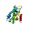
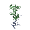
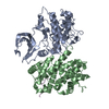
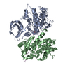
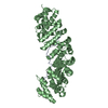
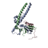
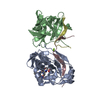
| Title | Crystal structure of the retinoblastoma tumour suppressor protein bound to E2F peptide | ||||||
 Components Components |
| ||||||
 Keywords Keywords | APOPTOSIS / TUMOUR SUPPRESSOR / CELL CYCLE REGULATION / DNA-BINDING | ||||||
| Function / homology |  Function and homology information Function and homology informationDefective translocation of RB1 mutants to the nucleus / enucleate erythrocyte differentiation / negative regulation of fat cell proliferation / Rb-E2F complex / regulation of lipid kinase activity / positive regulation of collagen fibril organization / lens fiber cell apoptotic process / negative regulation of myofibroblast differentiation / maintenance of mitotic sister chromatid cohesion / cell morphogenesis involved in neuron differentiation ...Defective translocation of RB1 mutants to the nucleus / enucleate erythrocyte differentiation / negative regulation of fat cell proliferation / Rb-E2F complex / regulation of lipid kinase activity / positive regulation of collagen fibril organization / lens fiber cell apoptotic process / negative regulation of myofibroblast differentiation / maintenance of mitotic sister chromatid cohesion / cell morphogenesis involved in neuron differentiation / chromatin lock complex / sister chromatid biorientation / importin-alpha family protein binding / positive regulation of transcription regulatory region DNA binding / Aberrant regulation of mitotic exit in cancer due to RB1 defects / positive regulation of extracellular matrix organization / Inhibition of replication initiation of damaged DNA by RB1/E2F1 / regulation of centromere complex assembly / positive regulation of macrophage differentiation / glial cell apoptotic process / tissue homeostasis / protein localization to chromosome, centromeric region / positive regulation of mitotic metaphase/anaphase transition / Transcription of E2F targets under negative control by p107 (RBL1) and p130 (RBL2) in complex with HDAC1 / negative regulation of hepatocyte apoptotic process / neuron maturation / mRNA stabilization / myoblast differentiation / Transcription of E2F targets under negative control by DREAM complex / Activation of NOXA and translocation to mitochondria / digestive tract development / anoikis / aortic valve morphogenesis / Replication of the SARS-CoV-1 genome / SWI/SNF complex / negative regulation of cold-induced thermogenesis / negative regulation of DNA binding / Activation of PUMA and translocation to mitochondria / negative regulation of glial cell proliferation / DNA-binding transcription activator activity / Formation of Senescence-Associated Heterochromatin Foci (SAHF) / smoothened signaling pathway / negative regulation of G1/S transition of mitotic cell cycle / hepatocyte apoptotic process / negative regulation of fat cell differentiation / G2 Phase / Phosphorylation of proteins involved in G1/S transition by active Cyclin E:Cdk2 complexes / G1/S-Specific Transcription / RUNX2 regulates osteoblast differentiation / Transcriptional Regulation by E2F6 / Defective binding of RB1 mutants to E2F1,(E2F2, E2F3) / skeletal muscle cell differentiation / regulation of G1/S transition of mitotic cell cycle / intrinsic apoptotic signaling pathway by p53 class mediator / negative regulation of apoptotic signaling pathway / negative regulation of cell cycle / chromosome organization / chondrocyte differentiation / TP53 Regulates Transcription of Genes Involved in G1 Cell Cycle Arrest / glial cell proliferation / Cyclin E associated events during G1/S transition / Cyclin A:Cdk2-associated events at S phase entry / cis-regulatory region sequence-specific DNA binding / forebrain development / Nuclear events stimulated by ALK signaling in cancer / striated muscle cell differentiation / regulation of mitotic cell cycle / Condensation of Prophase Chromosomes / DNA damage checkpoint signaling / epithelial cell proliferation / negative regulation of protein kinase activity / RNA polymerase II transcription regulatory region sequence-specific DNA binding / negative regulation of smoothened signaling pathway / negative regulation of DNA-binding transcription factor activity / phosphoprotein binding / APC/C:Cdh1 mediated degradation of Cdc20 and other APC/C:Cdh1 targeted proteins in late mitosis/early G1 / negative regulation of cell growth / PML body / Oncogene Induced Senescence / Pre-NOTCH Transcription and Translation / spindle / negative regulation of inflammatory response / kinase binding / G1/S transition of mitotic cell cycle / RNA polymerase II transcription regulator complex / cellular response to insulin stimulus / Transcriptional regulation of granulopoiesis / positive regulation of fibroblast proliferation / intrinsic apoptotic signaling pathway in response to DNA damage / negative regulation of epithelial cell proliferation / neuron projection development / transcription corepressor activity / Cyclin D associated events in G1 / sequence-specific double-stranded DNA binding / disordered domain specific binding / heterochromatin formation / cellular response to xenobiotic stimulus / MLL4 and MLL3 complexes regulate expression of PPARG target genes in adipogenesis and hepatic steatosis / Replication of the SARS-CoV-2 genome / Oxidative Stress Induced Senescence Similarity search - Function | ||||||
| Biological species |  HOMO SAPIENS (human) HOMO SAPIENS (human) | ||||||
| Method |  X-RAY DIFFRACTION / SYNCHROTRON / MOLECULAR REPLACEMENT / Resolution: 2.6 Å X-RAY DIFFRACTION / SYNCHROTRON / MOLECULAR REPLACEMENT / Resolution: 2.6 Å | ||||||
 Authors Authors | Xiao, B. / Spencer, J. / Clements, A. / Ali-Khan, N. / Mittnacht, S. / Broceno, C. / Burghammer, M. / Perrakis, A. / Marmorstein, R. / Gamblin, S.J. | ||||||
 Citation Citation | Journal: Proc.Natl.Acad.Sci.USA / Year: 2003 Title: Crystal Structure of the Retinoblastoma Tumor Suppressor Protein Bound to E2F and the Molecular Basis of its Regulation Authors: Xiao, B. / Spencer, J. / Clements, A. / Ali-Khan, N. / Mittnacht, S. / Broceno, C. / Burghammer, M. / Perrakis, A. / Marmorstein, R. / Gamblin, S.J. | ||||||
| History |
|
- Structure visualization
| Structure viewer | Molecule: MolmilJmol/JSmol |
|---|
- Downloads & links
-Download
| PDBx/mmCIF format | 1o9k.cif.gz | 296.1 KB | Display | PDBx/mmCIF format |
|---|---|---|---|---|
| PDB format | pdb1o9k.ent.gz | 243 KB | Display | PDB format |
| PDBx/mmJSON format | 1o9k.json.gz | Tree view | PDBx/mmJSON format | |
| Others |  Other downloads Other downloads |
-Validation report
| Arichive directory | https://data.pdbj.org/pub/pdb/validation_reports/o9/1o9kftp://data.pdbj.org/pub/pdb/validation_reports/o9/1o9k | HTTPS FTP |
|---|
-Related structure data
| Related structure data | 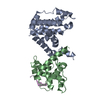 1guxS S: Starting model for refinement |
|---|---|
| Similar structure data |


-Links
 PDBj
PDBj


- Assembly
| Deposited unit | 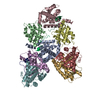
| ||||||||
|---|---|---|---|---|---|---|---|---|---|
| 1 | 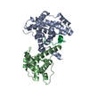
| ||||||||
| 2 | 
| ||||||||
| 3 | 
| ||||||||
| 4 | 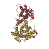
| ||||||||
| Unit cell |
| ||||||||
| Components on special symmetry positions |
|
-Components
| #1: Protein | Mass: 25100.910 Da / Num. of mol.: 4 / Fragment: DOMAIN A, RESIDUES 372-589 Source method: isolated from a genetically manipulated source Source: (gene. exp.) HOMO SAPIENS (human) / Production host:  #2: Protein | Mass: 18265.477 Da / Num. of mol.: 4 / Fragment: DOMAIN B, RESIDUES 636-787 Source method: isolated from a genetically manipulated source Source: (gene. exp.) HOMO SAPIENS (human) / Production host: #3: Protein/peptide | Mass: 2127.269 Da / Num. of mol.: 4 / Fragment: RESIDUES 409-426 / Source method: obtained synthetically / Source: (synth.) HOMO SAPIENS (human) / References: UniProt: Q01094#4: Water | ChemComp-HOH / |  Mass: 18.015 Da / Num. of mol.: 250 / Source method: isolated from a natural source / Formula: H2O Mass: 18.015 Da / Num. of mol.: 250 / Source method: isolated from a natural source / Formula: H2OCompound details | RETINOBLASTOMA-ASSOCIATED PROTEIN: REGULATOR OF OTHER GENES. ACTS AS A TUMOR SUPPRESSOR. INTERACTS ...RETINOBLAS | Has protein modification | Y | |
|---|
-Experimental details
-Experiment
| Experiment | Method: X-RAY DIFFRACTION / Number of used crystals: 1 |
|---|
- Sample preparation
| Crystal | Density Matthews: 2.45 Å3/Da / Density % sol: 49.84 % | ||||||||||||||||||||||||||||||||||||
|---|---|---|---|---|---|---|---|---|---|---|---|---|---|---|---|---|---|---|---|---|---|---|---|---|---|---|---|---|---|---|---|---|---|---|---|---|---|
| Crystal grow | pH: 7.8 / Details: pH 7.80 | ||||||||||||||||||||||||||||||||||||
| Crystal grow | *PLUS Temperature: 4 ℃ / Method: vapor diffusion, hanging drop | ||||||||||||||||||||||||||||||||||||
| Components of the solutions | *PLUS
|
-Data collection
| Diffraction | Mean temperature: 85 K |
|---|---|
| Diffraction source | Source: SYNCHROTRON / Site: ESRF  / Beamline: ID13 / Wavelength: 0.9 / Beamline: ID13 / Wavelength: 0.9 |
| Radiation | Protocol: SINGLE WAVELENGTH / Monochromatic (M) / Laue (L): M / Scattering type: x-ray |
| Radiation wavelength | Wavelength: 0.9 Å / Relative weight: 1 |
| Reflection | Resolution: 2.6→50 Å / Num. obs: 53846 / % possible obs: 88.1 % / Redundancy: 8.8 % / Rsym value: 0.094 / Net I/σ(I): 9.1 |
| Reflection shell | Resolution: 2.6→2.69 Å |
| Reflection | *PLUS Lowest resolution: 50 Å / Rmerge(I) obs: 0.094 |
| Reflection shell | *PLUS Rmerge(I) obs: 0.459 / Mean I/σ(I) obs: 2.5 |
- Processing
| Software |
| ||||||||||||||||||||||||||||||||||||||||||||||||||||||||||||||||||||||||||||||||||||||||||||||||||||||||||||||||||||||||||||||||||||||||||||||||||||||||||||||||||||||||||||||||||||||
|---|---|---|---|---|---|---|---|---|---|---|---|---|---|---|---|---|---|---|---|---|---|---|---|---|---|---|---|---|---|---|---|---|---|---|---|---|---|---|---|---|---|---|---|---|---|---|---|---|---|---|---|---|---|---|---|---|---|---|---|---|---|---|---|---|---|---|---|---|---|---|---|---|---|---|---|---|---|---|---|---|---|---|---|---|---|---|---|---|---|---|---|---|---|---|---|---|---|---|---|---|---|---|---|---|---|---|---|---|---|---|---|---|---|---|---|---|---|---|---|---|---|---|---|---|---|---|---|---|---|---|---|---|---|---|---|---|---|---|---|---|---|---|---|---|---|---|---|---|---|---|---|---|---|---|---|---|---|---|---|---|---|---|---|---|---|---|---|---|---|---|---|---|---|---|---|---|---|---|---|---|---|---|---|
| Refinement | Method to determine structure: MOLECULAR REPLACEMENT Starting model: PDB ENTRY 1GUX Resolution: 2.6→20 Å / Cor.coef. Fo:Fc: 0.89 / Cor.coef. Fo:Fc free: 0.823 / SU B: 13.486 / SU ML: 0.299 / TLS residual ADP flag: LIKELY RESIDUAL / Cross valid method: THROUGHOUT / ESU R Free: 0.406 / Stereochemistry target values: MAXIMUM LIKELIHOOD
| ||||||||||||||||||||||||||||||||||||||||||||||||||||||||||||||||||||||||||||||||||||||||||||||||||||||||||||||||||||||||||||||||||||||||||||||||||||||||||||||||||||||||||||||||||||||
| Solvent computation | Ion probe radii: 0.8 Å / Shrinkage radii: 0.8 Å / VDW probe radii: 1.4 Å / Solvent model: BABINET MODEL WITH MASK | ||||||||||||||||||||||||||||||||||||||||||||||||||||||||||||||||||||||||||||||||||||||||||||||||||||||||||||||||||||||||||||||||||||||||||||||||||||||||||||||||||||||||||||||||||||||
| Displacement parameters | Biso mean: 38.28 Å2
| ||||||||||||||||||||||||||||||||||||||||||||||||||||||||||||||||||||||||||||||||||||||||||||||||||||||||||||||||||||||||||||||||||||||||||||||||||||||||||||||||||||||||||||||||||||||
| Refinement step | Cycle: LAST / Resolution: 2.6→20 Å
| ||||||||||||||||||||||||||||||||||||||||||||||||||||||||||||||||||||||||||||||||||||||||||||||||||||||||||||||||||||||||||||||||||||||||||||||||||||||||||||||||||||||||||||||||||||||
| Refine LS restraints |
|