Defective translocation of RB1 mutants to the nucleus / enucleate erythrocyte differentiation / regulation of DNA biosynthetic process / Rb-E2F complex / negative regulation of fat cell proliferation / regulation of lipid kinase activity / lens fiber cell apoptotic process / positive regulation of collagen fibril organization / maintenance of mitotic sister chromatid cohesion / negative regulation of myofibroblast differentiation ...Defective translocation of RB1 mutants to the nucleus / enucleate erythrocyte differentiation / regulation of DNA biosynthetic process / Rb-E2F complex / negative regulation of fat cell proliferation / regulation of lipid kinase activity / lens fiber cell apoptotic process / positive regulation of collagen fibril organization / maintenance of mitotic sister chromatid cohesion / negative regulation of myofibroblast differentiation / chromatin lock complex / cell morphogenesis involved in neuron differentiation / Positive Regulation of CDH1 Gene Transcription / negative regulation of DNA binding / sister chromatid biorientation / positive regulation of transcription regulatory region DNA binding / positive regulation of extracellular matrix organization / Aberrant regulation of mitotic exit in cancer due to RB1 defects / Inhibition of replication initiation of damaged DNA by RB1/E2F1 / glial cell apoptotic process / positive regulation of macrophage differentiation / negative regulation of hepatocyte apoptotic process / protein localization to chromosome, centromeric region / tissue homeostasis / importin-alpha family protein binding / neuron maturation / Transcription of E2F targets under negative control by p107 (RBL1) and p130 (RBL2) in complex with HDAC1 / myoblast differentiation / positive regulation of mitotic metaphase/anaphase transition / cellular response to fatty acid / Transcription of E2F targets under negative control by DREAM complex / Activation of NOXA and translocation to mitochondria / quinolinate biosynthetic process / Replication of the SARS-CoV-1 genome / aortic valve morphogenesis / digestive tract development / anoikis / Activation of PUMA and translocation to mitochondria / negative regulation of cold-induced thermogenesis / negative regulation of glial cell proliferation / SWI/SNF complex / mRNA stabilization / Formation of Senescence-Associated Heterochromatin Foci (SAHF) / smoothened signaling pathway / DNA-binding transcription activator activity / G2 Phase / negative regulation of G1/S transition of mitotic cell cycle / Phosphorylation of proteins involved in G1/S transition by active Cyclin E:Cdk2 complexes / negative regulation of fat cell differentiation / G1/S-Specific Transcription / hepatocyte apoptotic process / nuclear chromosome / RUNX2 regulates osteoblast differentiation / forebrain development / Defective binding of RB1 mutants to E2F1,(E2F2, E2F3) / Transcriptional Regulation by E2F6 / negative regulation of cell cycle / intrinsic apoptotic signaling pathway by p53 class mediator / G0 and Early G1 / regulation of G1/S transition of mitotic cell cycle / : / negative regulation of apoptotic signaling pathway / epidermis development / positive regulation of glial cell proliferation / skeletal muscle cell differentiation / chondrocyte differentiation / chromosome organization / TP53 Regulates Transcription of Genes Involved in G1 Cell Cycle Arrest / negative regulation of protein kinase activity / Cyclin E associated events during G1/S transition / positive regulation of G1/S transition of mitotic cell cycle / Cyclin A:Cdk2-associated events at S phase entry / glial cell proliferation / cis-regulatory region sequence-specific DNA binding / striated muscle cell differentiation / Nuclear events stimulated by ALK signaling in cancer / epithelial cell proliferation / regulation of mitotic cell cycle / TP53 Regulates Transcription of Genes Involved in G2 Cell Cycle Arrest / DNA damage checkpoint signaling / Condensation of Prophase Chromosomes / RNA polymerase II transcription regulatory region sequence-specific DNA binding / negative regulation of smoothened signaling pathway / phosphoprotein binding / G1/S transition of mitotic cell cycle / cellular response to nerve growth factor stimulus / SMAD2/SMAD3:SMAD4 heterotrimer regulates transcription / negative regulation of cell growth / PML body / Oncogene Induced Senescence / APC/C:Cdh1 mediated degradation of Cdc20 and other APC/C:Cdh1 targeted proteins in late mitosis/early G1 / Pre-NOTCH Transcription and Translation / negative regulation of inflammatory response / cellular response to xenobiotic stimulus / RNA polymerase II transcription regulator complex / spindle / positive regulation of fibroblast proliferation / intrinsic apoptotic signaling pathway in response to DNA damage / negative regulation of epithelial cell proliferation / kinase binding Similarity search - Function
Single alpha-helices involved in coiled-coils or other helix-helix interfaces - #530 / Single alpha-helices involved in coiled-coils or other helix-helix interfaces - #540 / Transcription factor DP / Transcription factor DP, C-terminal / Transcription factor DP / Transcription factor DP, C-terminal domain superfamily / Transcription factor DP / Transcription factor DP / E2F transcription factor, CC-MB domain / E2F transcription factor CC-MB domain ...Single alpha-helices involved in coiled-coils or other helix-helix interfaces - #530 / Single alpha-helices involved in coiled-coils or other helix-helix interfaces - #540 / Transcription factor DP / Transcription factor DP, C-terminal / Transcription factor DP / Transcription factor DP, C-terminal domain superfamily / Transcription factor DP / Transcription factor DP / E2F transcription factor, CC-MB domain / E2F transcription factor CC-MB domain / E2F Family / E2F-DP heterodimerization region / E2F/DP family, winged-helix DNA-binding domain / E2F/DP family winged-helix DNA-binding domain / E2F/DP family winged-helix DNA-binding domain / Rb C-terminal domain / Retinoblastoma-associated protein, B-box / Retinoblastoma-associated protein, A-box / Retinoblastoma-associated protein, C-terminal / Retinoblastoma-associated protein, N-terminal / Retinoblastoma protein family / Retinoblastoma-associated protein B domain / Retinoblastoma-associated protein A domain / Domain of unknown function (DUF3452) / Domain of unknown function (DUF3452) / Retinoblastoma-associated protein A domain / Rb C-terminal domain / Butyryl-CoA Dehydrogenase, subunit A; domain 3 / Single alpha-helices involved in coiled-coils or other helix-helix interfaces / Cyclin-like / domain present in cyclins, TFIIB and Retinoblastoma / Cyclin-like superfamily / Helix non-globular / Special / Winged helix DNA-binding domain superfamily / Winged helix-like DNA-binding domain superfamily / Up-down Bundle / Mainly Alpha Similarity search - Domain/homology
In the structure databanks used in Yorodumi, some data are registered as the other names, "COVID-19 virus" and "2019-nCoV". Here are the details of the virus and the list of structure data.
Jan 31, 2019. EMDB accession codes are about to change! (news from PDBe EMDB page)
EMDB accession codes are about to change! (news from PDBe EMDB page)
The allocation of 4 digits for EMDB accession codes will soon come to an end. Whilst these codes will remain in use, new EMDB accession codes will include an additional digit and will expand incrementally as the available range of codes is exhausted. The current 4-digit format prefixed with “EMD-” (i.e. EMD-XXXX) will advance to a 5-digit format (i.e. EMD-XXXXX), and so on. It is currently estimated that the 4-digit codes will be depleted around Spring 2019, at which point the 5-digit format will come into force.
The EM Navigator/Yorodumi systems omit the EMD- prefix.
Related info.:Q: What is EMD? / ID/Accession-code notation in Yorodumi/EM Navigator
Yorodumi is a browser for structure data from EMDB, PDB, SASBDB, etc.
This page is also the successor to EM Navigator detail page, and also detail information page/front-end page for Omokage search.
The word "yorodu" (or yorozu) is an old Japanese word meaning "ten thousand". "mi" (miru) is to see.
Related info.:EMDB / PDB / SASBDB / Comparison of 3 databanks / Yorodumi Search / Aug 31, 2016. New EM Navigator & Yorodumi / Yorodumi Papers / Jmol/JSmol / Function and homology information / Changes in new EM Navigator and Yorodumi
 Movie
Movie Controller

 Yorodumi
 Open data
 Basic information
 Components
 Keywords
 Function and homology information
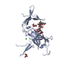
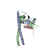
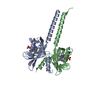
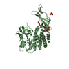
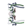
 Homo sapiens (human)
 X-RAY DIFFRACTION /
  Authors
 Citation
 Structure visualization
 Downloads & links
 Other downloads


 PDBj
PDBj


 Assembly
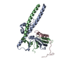

 Mass: 18.015 Da / Num. of mol.: 131 / Source method: isolated from a natural source / Formula: H2O
 Sample preparation
 Processing