 Movie
Movie Controller

[English] 日本語
 Yorodumi
- PDB-7a6p: Structural determinants underlying the adduct lifetime in a short... -
+ Open data

- Basic information
| Entry | Database: PDB / ID: 7a6p | ||||||
|---|---|---|---|---|---|---|---|
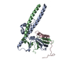
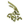
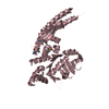
| Title | Structural determinants underlying the adduct lifetime in a short LOV protein PpSB2-LOV | ||||||
 Components Components | Putative Sensory box protein | ||||||
 Keywords Keywords | SIGNALING PROTEIN / LOV domain / PAS domain / Dimerization / Signaling blue light photoreceptor | ||||||
| Function / homology |  Function and homology information Function and homology information | ||||||
| Biological species |  Pseudomonas putida (bacteria) Pseudomonas putida (bacteria) | ||||||
| Method |  X-RAY DIFFRACTION / SYNCHROTRON / MOLECULAR REPLACEMENT / Resolution: 1.93 Å X-RAY DIFFRACTION / SYNCHROTRON / MOLECULAR REPLACEMENT / Resolution: 1.93 Å | ||||||
 Authors Authors | Arinkin, V. / Granzin, J. / Batra-Safferling, R. | ||||||
 Citation Citation | Journal: Febs J. / Year: 2021 Title: Structural determinants underlying the adduct lifetime in the LOV proteins of Pseudomonas putida. Authors: Arinkin, V. / Granzin, J. / Krauss, U. / Jaeger, K.E. / Willbold, D. / Batra-Safferling, R. | ||||||
| History |
|
- Structure visualization
| Structure viewer | Molecule: MolmilJmol/JSmol |
|---|
- Downloads & links
-Download
| PDBx/mmCIF format | 7a6p.cif.gz | 144.6 KB | Display | PDBx/mmCIF format |
|---|---|---|---|---|
| PDB format | pdb7a6p.ent.gz | 107.1 KB | Display | PDB format |
| PDBx/mmJSON format | 7a6p.json.gz | Tree view | PDBx/mmJSON format | |
| Others |  Other downloads Other downloads |
-Validation report
| Arichive directory | https://data.pdbj.org/pub/pdb/validation_reports/a6/7a6pftp://data.pdbj.org/pub/pdb/validation_reports/a6/7a6p | HTTPS FTP |
|---|
-Related structure data
| Related structure data | 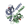 3sw1S S: Starting model for refinement |
|---|---|
| Similar structure data |


-Links
 PDBj
PDBj


- Assembly
| Deposited unit | 
| ||||||||||||
|---|---|---|---|---|---|---|---|---|---|---|---|---|---|
| 1 |
| ||||||||||||
| Unit cell |
|
-Components
| #1: Protein | Mass: 19210.469 Da / Num. of mol.: 2 Source method: isolated from a genetically manipulated source Source: (gene. exp.) Pseudomonas putida (strain ATCC 47054 / DSM 6125 / NCIMB 11950 / KT2440) (bacteria)Strain: ATCC 47054 / DSM 6125 / NCIMB 11950 / KT2440 / Gene: PP_2739 / Production host: #2: Chemical |   Mass: 106.120 Da / Num. of mol.: 2 / Source method: obtained synthetically / Formula: C4H10O3 / Feature type: SUBJECT OF INVESTIGATION Mass: 106.120 Da / Num. of mol.: 2 / Source method: obtained synthetically / Formula: C4H10O3 / Feature type: SUBJECT OF INVESTIGATION#3: Chemical |   Mass: 456.344 Da / Num. of mol.: 2 / Source method: obtained synthetically / Formula: C17H21N4O9P / Feature type: SUBJECT OF INVESTIGATION Mass: 456.344 Da / Num. of mol.: 2 / Source method: obtained synthetically / Formula: C17H21N4O9P / Feature type: SUBJECT OF INVESTIGATION#4: Water | ChemComp-HOH / |  Mass: 18.015 Da / Num. of mol.: 82 / Source method: isolated from a natural source / Formula: H2O Mass: 18.015 Da / Num. of mol.: 82 / Source method: isolated from a natural source / Formula: H2OHas ligand of interest | Y | |
|---|
-Experimental details
-Experiment
| Experiment | Method: X-RAY DIFFRACTION / Number of used crystals: 1 |
|---|
- Sample preparation
| Crystal | Density Matthews: 2.23 Å3/Da / Density % sol: 44.6 % / Description: needle |
|---|---|
| Crystal grow | Temperature: 292.15 K / Method: vapor diffusion, sitting drop / pH: 5.3 Details: Sodium acetate, polyethylene glycol 200, polyethylene glycol 3350 PH range: 4.9 - 5.3 |
-Data collection
| Diffraction | Mean temperature: 100 K / Serial crystal experiment: N | ||||||||||||||||||||||||
|---|---|---|---|---|---|---|---|---|---|---|---|---|---|---|---|---|---|---|---|---|---|---|---|---|---|
| Diffraction source | Source: SYNCHROTRON / Site: ESRF  / Beamline: ID23-2 / Wavelength: 0.8726 Å / Beamline: ID23-2 / Wavelength: 0.8726 Å | ||||||||||||||||||||||||
| Detector | Type: DECTRIS PILATUS3 X 2M / Detector: PIXEL / Date: Mar 8, 2015 | ||||||||||||||||||||||||
| Radiation | Protocol: SINGLE WAVELENGTH / Monochromatic (M) / Laue (L): M / Scattering type: x-ray | ||||||||||||||||||||||||
| Radiation wavelength | Wavelength: 0.8726 Å / Relative weight: 1 | ||||||||||||||||||||||||
| Reflection | Resolution: 1.93→44.88 Å / Num. obs: 25353 / % possible obs: 100 % / Redundancy: 4.2 % / CC1/2: 0.997 / Rmerge(I) obs: 0.097 / Rpim(I) all: 0.053 / Rrim(I) all: 0.111 / Net I/σ(I): 8.9 / Num. measured all: 107111 | ||||||||||||||||||||||||
| Reflection shell | Diffraction-ID: 1
|
- Processing
| Software |
| ||||||||||||||||||||||||
|---|---|---|---|---|---|---|---|---|---|---|---|---|---|---|---|---|---|---|---|---|---|---|---|---|---|
| Refinement | Method to determine structure: MOLECULAR REPLACEMENT Starting model: 3SW1 Resolution: 1.93→44.88 Å / Cross valid method: FREE R-VALUE Stereochemistry target values: GeoStd + Monomer Library + CDL v1.2
| ||||||||||||||||||||||||
| Displacement parameters | Biso mean: 39.18 Å2 | ||||||||||||||||||||||||
| Refinement step | Cycle: LAST / Resolution: 1.93→44.88 Å
| ||||||||||||||||||||||||
| Refine LS restraints |
|
