 Movie
Movie Controller


+ Open data

- Basic information
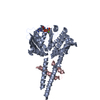
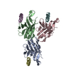
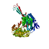
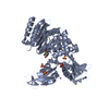
| Entry | Database: PDB / ID: 3qku | ||||||
|---|---|---|---|---|---|---|---|
| Title | Mre11 Rad50 binding domain in complex with Rad50 and AMP-PNP | ||||||
 Components Components |
| ||||||
 Keywords Keywords | REPLICATION / RecA-like fold / coiled-coils / ATPase / exonuclease / endonuclease / ATP binding / DNA binding | ||||||
| Function / homology |  Function and homology information Function and homology informationDNA end binding / Y-form DNA binding / DNA double-strand break processing / 3'-5' exonuclease activity / DNA endonuclease activity / manganese ion binding / double-strand break repair / endonuclease activity / Hydrolases; Acting on ester bonds / ATP hydrolysis activity ...DNA end binding / Y-form DNA binding / DNA double-strand break processing / 3'-5' exonuclease activity / DNA endonuclease activity / manganese ion binding / double-strand break repair / endonuclease activity / Hydrolases; Acting on ester bonds / ATP hydrolysis activity / zinc ion binding / ATP binding / identical protein binding Similarity search - Function | ||||||
| Biological species |   Pyrococcus furiosus (archaea) Pyrococcus furiosus (archaea) | ||||||
| Method |  X-RAY DIFFRACTION / SYNCHROTRON / MOLECULAR REPLACEMENT / Resolution: 3.3 Å X-RAY DIFFRACTION / SYNCHROTRON / MOLECULAR REPLACEMENT / Resolution: 3.3 Å | ||||||
 Authors Authors | Williams, G.J. / Williams, R.S. / Arvai, A. / Moncalian, G. / Tainer, J.A. | ||||||
 Citation Citation | Journal: Nat.Struct.Mol.Biol. / Year: 2011 Title: ABC ATPase signature helices in Rad50 link nucleotide state to Mre11 interface for DNA repair. Authors: Williams, G.J. / Williams, R.S. / Williams, J.S. / Moncalian, G. / Arvai, A.S. / Limbo, O. / Guenther, G. / Sildas, S. / Hammel, M. / Russell, P. / Tainer, J.A. | ||||||
| History |
|
- Structure visualization
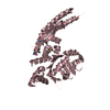
| Structure viewer | Molecule: MolmilJmol/JSmol |
|---|
- Downloads & links
-Download
| PDBx/mmCIF format | 3qku.cif.gz | 156.2 KB | Display | PDBx/mmCIF format |
|---|---|---|---|---|
| PDB format | pdb3qku.ent.gz | 121.8 KB | Display | PDB format |
| PDBx/mmJSON format | 3qku.json.gz | Tree view | PDBx/mmJSON format | |
| Others |  Other downloads Other downloads |
-Validation report
| Arichive directory | https://data.pdbj.org/pub/pdb/validation_reports/qk/3qkuftp://data.pdbj.org/pub/pdb/validation_reports/qk/3qku | HTTPS FTP |
|---|
-Related structure data
| Related structure data |  3qkrC  3qksC  3qktC  1us8S C: citing same article ( S: Starting model for refinement |
|---|---|
| Similar structure data |
-Links
 PDBj
PDBj


- Assembly
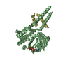
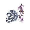
| Deposited unit | 
| ||||||||
|---|---|---|---|---|---|---|---|---|---|
| 1 | 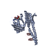
| ||||||||
| 2 | 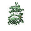
| ||||||||
| Unit cell |
|
-Components
| #1: Protein | Mass: 41063.473 Da / Num. of mol.: 2 / Fragment: UNP residues 1-187, 716-882 Source method: isolated from a genetically manipulated source Source: (gene. exp.) Pyrococcus furiosus (archaea) / Gene: rad50, PF1167 / Production host:  References: UniProt: P58301, Hydrolases; Acting on acid anhydrides #2: Protein/peptide | | Mass: 4062.568 Da / Num. of mol.: 1 / Fragment: Rad50 binding domain (UNP residues 348-381) Source method: isolated from a genetically manipulated source Source: (gene. exp.) Pyrococcus furiosus (archaea) / Gene: mre11, PF1166 / Production host: References: UniProt: Q8U1N9, Hydrolases; Acting on ester bonds #3: Chemical |   Mass: 506.196 Da / Num. of mol.: 2 / Source method: obtained synthetically / Formula: C10H17N6O12P3 / Comment: AMP-PNP, energy-carrying molecule analogue*YM Mass: 506.196 Da / Num. of mol.: 2 / Source method: obtained synthetically / Formula: C10H17N6O12P3 / Comment: AMP-PNP, energy-carrying molecule analogue*YM#4: Chemical |   Mass: 24.305 Da / Num. of mol.: 2 / Source method: obtained synthetically / Formula: Mg Mass: 24.305 Da / Num. of mol.: 2 / Source method: obtained synthetically / Formula: MgSequence details | THE RAD50 ABC-ATPASE CONSTRUCT CONSISTS OF UNP RESIDUES 1-187 AND 716-882 CONNECTED BY AN ...THE RAD50 ABC-ATPASE CONSTRUCT CONSISTS OF UNP RESIDUES 1-187 AND 716-882 CONNECTED BY AN ENGINEERED | |
|---|
-Experimental details
-Experiment
| Experiment | Method: X-RAY DIFFRACTION / Number of used crystals: 1 |
|---|
- Sample preparation
| Crystal | Density Matthews: 3.44 Å3/Da / Density % sol: 64.21 % |
|---|---|
| Crystal grow | Temperature: 293 K / Method: vapor diffusion, hanging drop / pH: 8.5 Details: 100 mM Tris, 200-300 mM lithium sulfate, 12-13% PEG3350, pH 8.5, VAPOR DIFFUSION, HANGING DROP, temperature 293K |
-Data collection
| Diffraction | Mean temperature: 100 K |
|---|---|
| Diffraction source | Source: SYNCHROTRON / Site: ALS  / Beamline: 12.3.1 / Wavelength: 1.11583 / Beamline: 12.3.1 / Wavelength: 1.11583 |
| Detector | Type: ADSC QUANTUM 315 / Detector: CCD / Date: Nov 1, 2007 |
| Radiation | Monochromator: Kohzu Dual Double Crystal Monochromator (DDCM) Si(111) Protocol: SINGLE WAVELENGTH / Monochromatic (M) / Laue (L): M / Scattering type: x-ray |
| Radiation wavelength | Wavelength: 1.11583 Å / Relative weight: 1 |
| Reflection | Resolution: 3.3→153.6 Å / Num. all: 18722 / Num. obs: 18667 / % possible obs: 99.7 % / Observed criterion σ(F): -3 / Observed criterion σ(I): -3 / Redundancy: 9.8 % / Rmerge(I) obs: 0.048 / Net I/σ(I): 35.7 |
| Reflection shell | Resolution: 3.301→3.387 Å / Redundancy: 10.1 % / Rmerge(I) obs: 0.66 / Mean I/σ(I) obs: 3.6 / % possible all: 99.41 |
- Processing
| Software |
| ||||||||||||||||||||||||||||||||||||||||||||||||||||||||||||||||||||||||||||||||||||||||||||||||||||||||||||||||||||||||||||||||||||||||||||||||||||||||||||||||||||||||||
|---|---|---|---|---|---|---|---|---|---|---|---|---|---|---|---|---|---|---|---|---|---|---|---|---|---|---|---|---|---|---|---|---|---|---|---|---|---|---|---|---|---|---|---|---|---|---|---|---|---|---|---|---|---|---|---|---|---|---|---|---|---|---|---|---|---|---|---|---|---|---|---|---|---|---|---|---|---|---|---|---|---|---|---|---|---|---|---|---|---|---|---|---|---|---|---|---|---|---|---|---|---|---|---|---|---|---|---|---|---|---|---|---|---|---|---|---|---|---|---|---|---|---|---|---|---|---|---|---|---|---|---|---|---|---|---|---|---|---|---|---|---|---|---|---|---|---|---|---|---|---|---|---|---|---|---|---|---|---|---|---|---|---|---|---|---|---|---|---|---|---|---|
| Refinement | Method to determine structure: MOLECULAR REPLACEMENT Starting model: PDB ENTRY 1US8 Resolution: 3.3→50 Å / Cor.coef. Fo:Fc: 0.936 / Cor.coef. Fo:Fc free: 0.909 / SU B: 32.231 / SU ML: 0.511 / Cross valid method: THROUGHOUT / σ(F): 0 / ESU R Free: 0.584 / Stereochemistry target values: MAXIMUM LIKELIHOOD / Details: HYDROGENS HAVE BEEN ADDED IN THE RIDING POSITIONS
| ||||||||||||||||||||||||||||||||||||||||||||||||||||||||||||||||||||||||||||||||||||||||||||||||||||||||||||||||||||||||||||||||||||||||||||||||||||||||||||||||||||||||||
| Solvent computation | Ion probe radii: 0.8 Å / Shrinkage radii: 0.8 Å / VDW probe radii: 1.25 Å / Solvent model: BABINET MODEL WITH MASK | ||||||||||||||||||||||||||||||||||||||||||||||||||||||||||||||||||||||||||||||||||||||||||||||||||||||||||||||||||||||||||||||||||||||||||||||||||||||||||||||||||||||||||
| Displacement parameters | Biso mean: 134.928 Å2
| ||||||||||||||||||||||||||||||||||||||||||||||||||||||||||||||||||||||||||||||||||||||||||||||||||||||||||||||||||||||||||||||||||||||||||||||||||||||||||||||||||||||||||
| Refinement step | Cycle: LAST / Resolution: 3.3→50 Å
| ||||||||||||||||||||||||||||||||||||||||||||||||||||||||||||||||||||||||||||||||||||||||||||||||||||||||||||||||||||||||||||||||||||||||||||||||||||||||||||||||||||||||||
| Refine LS restraints |
| ||||||||||||||||||||||||||||||||||||||||||||||||||||||||||||||||||||||||||||||||||||||||||||||||||||||||||||||||||||||||||||||||||||||||||||||||||||||||||||||||||||||||||
| LS refinement shell | Resolution: 3.301→3.387 Å / Total num. of bins used: 20
|
