 Movie
Movie Controller

[English] 日本語
 Yorodumi
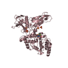
- PDB-2vgk: Crystal structure of Actinomadura R39 DD-peptidase complexed with... -
+ Open data

- Basic information
| Entry | Database: PDB / ID: 2vgk | ||||||
|---|---|---|---|---|---|---|---|
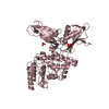
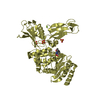
| Title | Crystal structure of Actinomadura R39 DD-peptidase complexed with a peptidoglycan-mimetic cephalosporin | ||||||
 Components Components | D-ALANYL-D-ALANINE CARBOXYPEPTIDASE | ||||||
 Keywords Keywords | HYDROLASE / CELL WALL BIOGENESIS/DEGRADATION / ANTIBIOTIC RESISTANCE / PEPTIDOGLYCAN SYNTHESIS / PROTEASE / SECRETED / CELL SHAPE / CARBOXYPEPTIDASE / PENICILLIN-BINDING / ACTINOMADURA / PEPTIDOGLYCAN / TRANSPEPTIDASE | ||||||
| Function / homology |  Function and homology information Function and homology informationserine-type D-Ala-D-Ala carboxypeptidase / serine-type D-Ala-D-Ala carboxypeptidase activity / peptidoglycan biosynthetic process / cell wall organization / regulation of cell shape / response to antibiotic / proteolysis / extracellular region Similarity search - Function | ||||||
| Biological species |  ACTINOMADURA SP. (bacteria) ACTINOMADURA SP. (bacteria) | ||||||
| Method |  X-RAY DIFFRACTION / MOLECULAR REPLACEMENT / Resolution: 2.25 Å X-RAY DIFFRACTION / MOLECULAR REPLACEMENT / Resolution: 2.25 Å | ||||||
 Authors Authors | Sauvage, E. / kerff, F. / Herman, R. / Charlier, P. | ||||||
 Citation Citation | Journal: J.Mol.Biol. / Year: 2008 Title: Crystal Structures of Complexes of Bacterial Dd-Peptidases with Peptidoglycan-Mimetic Ligands: The Substrate Specificity Puzzle. Authors: Sauvage, E. / Powell, A.J. / Heilemann, J. / Josephin, H.R. / Charlier, P. / Davies, C. / Pratt, R.F. | ||||||
| History |
| ||||||
| Remark 700 | SHEET THE SHEET STRUCTURE OF THIS MOLECULE IS BIFURCATED. IN ORDER TO REPRESENT THIS FEATURE IN ... SHEET THE SHEET STRUCTURE OF THIS MOLECULE IS BIFURCATED. IN ORDER TO REPRESENT THIS FEATURE IN THE SHEET RECORDS BELOW, TWO SHEETS ARE DEFINED. |
- Structure visualization
| Structure viewer | Molecule: MolmilJmol/JSmol |
|---|
- Downloads & links
-Download
| PDBx/mmCIF format | 2vgk.cif.gz | 346.4 KB | Display | PDBx/mmCIF format |
|---|---|---|---|---|
| PDB format | pdb2vgk.ent.gz | 282.6 KB | Display | PDB format |
| PDBx/mmJSON format | 2vgk.json.gz | Tree view | PDBx/mmJSON format | |
| Others |  Other downloads Other downloads |
-Validation report
| Arichive directory | https://data.pdbj.org/pub/pdb/validation_reports/vg/2vgkftp://data.pdbj.org/pub/pdb/validation_reports/vg/2vgk | HTTPS FTP |
|---|
-Related structure data
| Related structure data |  2vgjC  3bebC  3becC 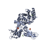 1w79S S: Starting model for refinement C: citing same article ( |
|---|---|
| Similar structure data |


-Links
 PDBj
PDBj
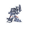
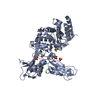
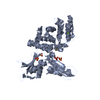
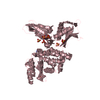
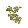
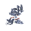
- Assembly
| Deposited unit | 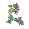
| ||||||||
|---|---|---|---|---|---|---|---|---|---|
| 1 | 
| ||||||||
| 2 | 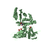
| ||||||||
| 3 | 
| ||||||||
| 4 | 
| ||||||||
| Unit cell |
|
-Components
| #1: Protein | Mass: 50083.812 Da / Num. of mol.: 4 / Source method: isolated from a natural source / Source: (natural) ACTINOMADURA SP. (bacteria) / Strain: R39References: UniProt: P39045, serine-type D-Ala-D-Ala carboxypeptidase #2: Chemical | 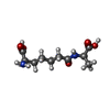  Mass: 246.260 Da / Num. of mol.: 3 / Source method: obtained synthetically / Formula: C10H18N2O5 Mass: 246.260 Da / Num. of mol.: 3 / Source method: obtained synthetically / Formula: C10H18N2O5#3: Chemical | ChemComp-SO4 /   Mass: 96.063 Da / Num. of mol.: 20 / Source method: obtained synthetically / Formula: SO4 Mass: 96.063 Da / Num. of mol.: 20 / Source method: obtained synthetically / Formula: SO4#4: Chemical | ChemComp-MG /   Mass: 24.305 Da / Num. of mol.: 4 / Source method: obtained synthetically / Formula: Mg Mass: 24.305 Da / Num. of mol.: 4 / Source method: obtained synthetically / Formula: Mg#5: Water | ChemComp-HOH / |  Mass: 18.015 Da / Num. of mol.: 495 / Source method: isolated from a natural source / Formula: H2O Mass: 18.015 Da / Num. of mol.: 495 / Source method: isolated from a natural source / Formula: H2O |
|---|
-Experimental details
-Experiment
| Experiment | Method: X-RAY DIFFRACTION / Number of used crystals: 1 |
|---|
- Sample preparation
| Crystal | Density Matthews: 2.71 Å3/Da / Density % sol: 54.3 % / Description: NONE |
|---|---|
| Crystal grow | pH: 8 / Details: pH 8 |
-Data collection
| Diffraction | Mean temperature: 100 K |
|---|---|
| Diffraction source | Source: ROTATING ANODE / Type: RIGAKU RU200 / Wavelength: 1.5418 |
| Detector | Type: MARRESEARCH / Detector: IMAGE PLATE / Date: Dec 1, 2004 / Details: MIRRORS |
| Radiation | Protocol: SINGLE WAVELENGTH / Monochromatic (M) / Laue (L): M / Scattering type: x-ray |
| Radiation wavelength | Wavelength: 1.5418 Å / Relative weight: 1 |
| Reflection | Resolution: 2.25→20 Å / Num. obs: 99513 / % possible obs: 99.1 % / Observed criterion σ(I): 0 / Redundancy: 3.7 % / Rmerge(I) obs: 0.17 / Net I/σ(I): 9.7 |
| Reflection shell | Resolution: 2.25→2.37 Å / Redundancy: 3.5 % / Rmerge(I) obs: 0.71 / Mean I/σ(I) obs: 2.3 / % possible all: 94.9 |
- Processing
| Software |
| ||||||||||||||||||||||||||||||||||||||||||||||||||||||||||||||||||||||||||||||||||||||||||||||||||||||||||||||||||||||||||||||||||||||||||||||||||||||||||||||||||||||||||||||||||||||
|---|---|---|---|---|---|---|---|---|---|---|---|---|---|---|---|---|---|---|---|---|---|---|---|---|---|---|---|---|---|---|---|---|---|---|---|---|---|---|---|---|---|---|---|---|---|---|---|---|---|---|---|---|---|---|---|---|---|---|---|---|---|---|---|---|---|---|---|---|---|---|---|---|---|---|---|---|---|---|---|---|---|---|---|---|---|---|---|---|---|---|---|---|---|---|---|---|---|---|---|---|---|---|---|---|---|---|---|---|---|---|---|---|---|---|---|---|---|---|---|---|---|---|---|---|---|---|---|---|---|---|---|---|---|---|---|---|---|---|---|---|---|---|---|---|---|---|---|---|---|---|---|---|---|---|---|---|---|---|---|---|---|---|---|---|---|---|---|---|---|---|---|---|---|---|---|---|---|---|---|---|---|---|---|
| Refinement | Method to determine structure: MOLECULAR REPLACEMENT Starting model: PDB ENTRY 1W79 Resolution: 2.25→15.71 Å / Cor.coef. Fo:Fc: 0.919 / Cor.coef. Fo:Fc free: 0.867 / SU B: 9.762 / SU ML: 0.23 / Cross valid method: THROUGHOUT / ESU R: 0.32 / ESU R Free: 0.253 / Stereochemistry target values: MAXIMUM LIKELIHOOD / Details: HYDROGENS HAVE BEEN ADDED IN THE RIDING POSITIONS.
| ||||||||||||||||||||||||||||||||||||||||||||||||||||||||||||||||||||||||||||||||||||||||||||||||||||||||||||||||||||||||||||||||||||||||||||||||||||||||||||||||||||||||||||||||||||||
| Solvent computation | Ion probe radii: 0.8 Å / Shrinkage radii: 0.8 Å / VDW probe radii: 1.4 Å / Solvent model: MASK | ||||||||||||||||||||||||||||||||||||||||||||||||||||||||||||||||||||||||||||||||||||||||||||||||||||||||||||||||||||||||||||||||||||||||||||||||||||||||||||||||||||||||||||||||||||||
| Displacement parameters | Biso mean: 26.41 Å2
| ||||||||||||||||||||||||||||||||||||||||||||||||||||||||||||||||||||||||||||||||||||||||||||||||||||||||||||||||||||||||||||||||||||||||||||||||||||||||||||||||||||||||||||||||||||||
| Refinement step | Cycle: LAST / Resolution: 2.25→15.71 Å
| ||||||||||||||||||||||||||||||||||||||||||||||||||||||||||||||||||||||||||||||||||||||||||||||||||||||||||||||||||||||||||||||||||||||||||||||||||||||||||||||||||||||||||||||||||||||
| Refine LS restraints |
|