 Movie
Movie Controller

[English] 日本語
 Yorodumi
- PDB-1us8: The Rad50 signature motif: essential to ATP binding and biologica... -
+ Open data

- Basic information
| Entry | Database: PDB / ID: 1us8 | ||||||
|---|---|---|---|---|---|---|---|
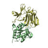
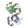
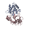
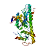
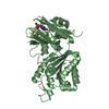
| Title | The Rad50 signature motif: essential to ATP binding and biological function | ||||||
 Components Components | (DNA DOUBLE-STRAND BREAK REPAIR RAD50 ATPASE) x 2 | ||||||
 Keywords Keywords | DNA REPAIR / ABC ATPASE / SIGNATURE MOTIF | ||||||
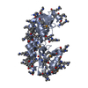
| Function / homology |  Function and homology information Function and homology informationdouble-strand break repair / ATP hydrolysis activity / zinc ion binding / ATP binding / identical protein binding Similarity search - Function | ||||||
| Biological species |   PYROCOCCUS FURIOSUS (archaea) PYROCOCCUS FURIOSUS (archaea) | ||||||
| Method |  X-RAY DIFFRACTION / SYNCHROTRON / MOLECULAR REPLACEMENT / Resolution: 2.1 Å X-RAY DIFFRACTION / SYNCHROTRON / MOLECULAR REPLACEMENT / Resolution: 2.1 Å | ||||||
 Authors Authors | Moncalian, G. / Lengsfeld, B. / Bhaskara, V. / Hopfner, K.P. / Karcher, A. / Alden, E. / Tainer, J.A. / Paull, T.T. | ||||||
 Citation Citation | Journal: J.Mol.Biol. / Year: 2004 Title: The Rad50 Signature Motif: Essential to ATP Binding and Biological Function Authors: Moncalian, G. / Lengsfeld, B. / Bhaskara, V. / Hopfner, K.P. / Karcher, A. / Alden, E. / Tainer, J.A. / Paull, T.T. | ||||||
| History |
|
- Structure visualization
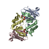
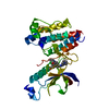
| Structure viewer | Molecule: MolmilJmol/JSmol |
|---|
- Downloads & links
-Download
| PDBx/mmCIF format | 1us8.cif.gz | 67.8 KB | Display | PDBx/mmCIF format |
|---|---|---|---|---|
| PDB format | pdb1us8.ent.gz | 49.8 KB | Display | PDB format |
| PDBx/mmJSON format | 1us8.json.gz | Tree view | PDBx/mmJSON format | |
| Others |  Other downloads Other downloads |
-Validation report
| Arichive directory | https://data.pdbj.org/pub/pdb/validation_reports/us/1us8ftp://data.pdbj.org/pub/pdb/validation_reports/us/1us8 | HTTPS FTP |
|---|
-Related structure data
| Related structure data | 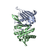 1f2tS S: Starting model for refinement |
|---|---|
| Similar structure data |


-Links
 PDBj
PDBj- Assembly
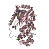
| Deposited unit | 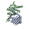
| ||||||||
|---|---|---|---|---|---|---|---|---|---|
| 1 |
| ||||||||
| Unit cell |
|
-Components
| #1: Protein | Mass: 16894.648 Da / Num. of mol.: 1 / Fragment: N-TERMINAL DOMAIN, RESIDUES 1-147 Source method: isolated from a genetically manipulated source Source: (gene. exp.) PYROCOCCUS FURIOSUS (archaea) / Plasmid: PET28 / Production host:  |
|---|---|
| #2: Protein | Mass: 16364.932 Da / Num. of mol.: 1 / Fragment: C-TERMINAL DOMAIN, RESIDUES 739-882 / Mutation: YES Source method: isolated from a genetically manipulated source Source: (gene. exp.) PYROCOCCUS FURIOSUS (archaea) / Plasmid: PET28 / Production host: |
| #3: Water | ChemComp-HOH /  Mass: 18.015 Da / Num. of mol.: 93 / Source method: isolated from a natural source / Formula: H2O Mass: 18.015 Da / Num. of mol.: 93 / Source method: isolated from a natural source / Formula: H2O |
| Compound details | ENGINEERED |
-Experimental details
-Experiment
| Experiment | Method: X-RAY DIFFRACTION / Number of used crystals: 1 |
|---|
- Sample preparation
| Crystal | Density Matthews: 2.43 Å3/Da / Density % sol: 48.94 % | |||||||||||||||||||||||||||||||||||||||||||||||||||||||||||||||||||||||||||||
|---|---|---|---|---|---|---|---|---|---|---|---|---|---|---|---|---|---|---|---|---|---|---|---|---|---|---|---|---|---|---|---|---|---|---|---|---|---|---|---|---|---|---|---|---|---|---|---|---|---|---|---|---|---|---|---|---|---|---|---|---|---|---|---|---|---|---|---|---|---|---|---|---|---|---|---|---|---|---|
| Crystal grow | pH: 7.5 Details: 20 % PEG8000, 0.1 M MES PH 6.0, 0.2 M CALCIUM ACETATE | |||||||||||||||||||||||||||||||||||||||||||||||||||||||||||||||||||||||||||||
| Crystal grow | *PLUS pH: 7 / Method: vapor diffusion, sitting drop | |||||||||||||||||||||||||||||||||||||||||||||||||||||||||||||||||||||||||||||
| Components of the solutions | *PLUS
|
-Data collection
| Diffraction | Mean temperature: 294 K |
|---|---|
| Diffraction source | Source: SYNCHROTRON / Site: SSRL  / Beamline: BL11-1 / Wavelength: 0.849961 / Beamline: BL11-1 / Wavelength: 0.849961 |
| Detector | Date: Nov 15, 2001 |
| Radiation | Protocol: SINGLE WAVELENGTH / Monochromatic (M) / Laue (L): M / Scattering type: x-ray |
| Radiation wavelength | Wavelength: 0.849961 Å / Relative weight: 1 |
| Reflection | Resolution: 2.1→19.75 Å / Num. obs: 25989 / % possible obs: 99.2 % / Observed criterion σ(I): 2 / Redundancy: 4 % / Biso Wilson estimate: 19.5 Å2 / Rmerge(I) obs: 0.057 / Net I/σ(I): 10 |
| Reflection shell | Resolution: 2.05→2.14 Å / Redundancy: 4 % / Rmerge(I) obs: 0.279 / Mean I/σ(I) obs: 3 / % possible all: 99.9 |
| Reflection | *PLUS Highest resolution: 2.1 Å / Lowest resolution: 20 Å / Num. measured all: 200189 / Rmerge(I) obs: 0.057 |
| Reflection shell | *PLUS % possible obs: 98.9 % / Rmerge(I) obs: 0.279 |
- Processing
| Software |
| ||||||||||||||||||||||||||||||||||||||||||||||||||||||||||||||||||||||||||||||||
|---|---|---|---|---|---|---|---|---|---|---|---|---|---|---|---|---|---|---|---|---|---|---|---|---|---|---|---|---|---|---|---|---|---|---|---|---|---|---|---|---|---|---|---|---|---|---|---|---|---|---|---|---|---|---|---|---|---|---|---|---|---|---|---|---|---|---|---|---|---|---|---|---|---|---|---|---|---|---|---|---|---|
| Refinement | Method to determine structure: MOLECULAR REPLACEMENT Starting model: PDB ENTRY 1F2T Resolution: 2.1→19.75 Å / Rfactor Rfree error: 0.007 / Data cutoff high absF: 407016.46 / Isotropic thermal model: RESTRAINED / Cross valid method: THROUGHOUT / σ(F): 0
| ||||||||||||||||||||||||||||||||||||||||||||||||||||||||||||||||||||||||||||||||
| Solvent computation | Solvent model: FLAT MODEL / ksol: 0.381882 e/Å3 | ||||||||||||||||||||||||||||||||||||||||||||||||||||||||||||||||||||||||||||||||
| Displacement parameters | Biso mean: 36 Å2
| ||||||||||||||||||||||||||||||||||||||||||||||||||||||||||||||||||||||||||||||||
| Refine analyze |
| ||||||||||||||||||||||||||||||||||||||||||||||||||||||||||||||||||||||||||||||||
| Refinement step | Cycle: LAST / Resolution: 2.1→19.75 Å
| ||||||||||||||||||||||||||||||||||||||||||||||||||||||||||||||||||||||||||||||||
| Refine LS restraints |
| ||||||||||||||||||||||||||||||||||||||||||||||||||||||||||||||||||||||||||||||||
| LS refinement shell | Resolution: 2.1→2.23 Å / Rfactor Rfree error: 0.018 / Total num. of bins used: 6
| ||||||||||||||||||||||||||||||||||||||||||||||||||||||||||||||||||||||||||||||||
| Xplor file |
| ||||||||||||||||||||||||||||||||||||||||||||||||||||||||||||||||||||||||||||||||
| Refinement | *PLUS Highest resolution: 2.1 Å / Lowest resolution: 20 Å / Num. reflection obs: 18606 / Num. reflection Rfree: 908 / % reflection Rfree: 5 % / Rfactor Rfree: 0.249 / Rfactor Rwork: 0.22 | ||||||||||||||||||||||||||||||||||||||||||||||||||||||||||||||||||||||||||||||||
| Solvent computation | *PLUS | ||||||||||||||||||||||||||||||||||||||||||||||||||||||||||||||||||||||||||||||||
| Displacement parameters | *PLUS | ||||||||||||||||||||||||||||||||||||||||||||||||||||||||||||||||||||||||||||||||
| Refine LS restraints | *PLUS
|
