 Movie
Movie Controller


+ Open data

- Basic information
| Entry | Database: PDB / ID: 1ii8 | ||||||
|---|---|---|---|---|---|---|---|
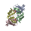
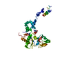
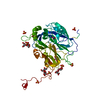
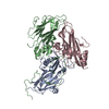
| Title | Crystal structure of the P. furiosus Rad50 ATPase domain | ||||||
 Components Components | (Rad50 ABC-ATPase) x 2 | ||||||
 Keywords Keywords | REPLICATION / Rad50 / Mre11 / DNA double-strand break repair / ATP | ||||||
| Function / homology |  Function and homology information Function and homology informationdouble-strand break repair / ATP hydrolysis activity / zinc ion binding / ATP binding / identical protein binding Similarity search - Function | ||||||
| Biological species |   Pyrococcus furiosus (archaea) Pyrococcus furiosus (archaea) | ||||||
| Method |  X-RAY DIFFRACTION / SYNCHROTRON / MOLECULAR REPLACEMENT / Resolution: 3.02 Å X-RAY DIFFRACTION / SYNCHROTRON / MOLECULAR REPLACEMENT / Resolution: 3.02 Å | ||||||
 Authors Authors | Hopfner, K.-P. / Karcher, A. / Craig, L. / Woo, T.T. / Carney, J.P. / Tainer, J.A. | ||||||
 Citation Citation | Journal: Cell(Cambridge,Mass.) / Year: 2001 Title: Structural biochemistry and interaction architecture of the DNA double-strand break repair Mre11 nuclease and Rad50-ATPase. Authors: Hopfner, K.P. / Karcher, A. / Craig, L. / Woo, T.T. / Carney, J.P. / Tainer, J.A. | ||||||
| History |
|
- Structure visualization
| Structure viewer | Molecule: MolmilJmol/JSmol |
|---|
- Downloads & links
-Download
| PDBx/mmCIF format | 1ii8.cif.gz | 91.2 KB | Display | PDBx/mmCIF format |
|---|---|---|---|---|
| PDB format | pdb1ii8.ent.gz | 67.8 KB | Display | PDB format |
| PDBx/mmJSON format | 1ii8.json.gz | Tree view | PDBx/mmJSON format | |
| Others |  Other downloads Other downloads |
-Validation report
| Arichive directory | https://data.pdbj.org/pub/pdb/validation_reports/ii/1ii8ftp://data.pdbj.org/pub/pdb/validation_reports/ii/1ii8 | HTTPS FTP |
|---|
-Related structure data
| Related structure data |  1ii7C 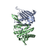 1f2tS C: citing same article ( S: Starting model for refinement |
|---|---|
| Similar structure data |


-Links
 PDBj
PDBj- Assembly
| Deposited unit | 
| ||||||||
|---|---|---|---|---|---|---|---|---|---|
| 1 |
| ||||||||
| 2 | 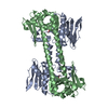
| ||||||||
| Unit cell |
|
-Components
| #1: Protein | Mass: 22672.248 Da / Num. of mol.: 1 / Fragment: N-terminal fragment Source method: isolated from a genetically manipulated source Source: (gene. exp.) Pyrococcus furiosus (archaea) / Production host:  | ||
|---|---|---|---|
| #2: Protein | Mass: 19981.195 Da / Num. of mol.: 1 / Fragment: C-terminal fragment Source method: isolated from a genetically manipulated source Source: (gene. exp.) Pyrococcus furiosus (archaea) / Production host: | ||
| #3: Chemical |   Mass: 94.971 Da / Num. of mol.: 2 / Source method: obtained synthetically / Formula: PO4 Mass: 94.971 Da / Num. of mol.: 2 / Source method: obtained synthetically / Formula: PO4#4: Water | ChemComp-HOH / |  Mass: 18.015 Da / Num. of mol.: 63 / Source method: isolated from a natural source / Formula: H2O Mass: 18.015 Da / Num. of mol.: 63 / Source method: isolated from a natural source / Formula: H2O |
-Experimental details
-Experiment
| Experiment | Method: X-RAY DIFFRACTION / Number of used crystals: 1 |
|---|
- Sample preparation
| Crystal | Density Matthews: 3.9 Å3/Da / Density % sol: 68.47 % | ||||||||||||||||||||||||||||||||||||||||||||||||||||||
|---|---|---|---|---|---|---|---|---|---|---|---|---|---|---|---|---|---|---|---|---|---|---|---|---|---|---|---|---|---|---|---|---|---|---|---|---|---|---|---|---|---|---|---|---|---|---|---|---|---|---|---|---|---|---|---|
| Crystal grow | Temperature: 300 K / Method: vapor diffusion, sitting drop / pH: 6.2 Details: 100 mM Na-Acetate, 8% PEG 6K, 10 mM Ca-Acetate, pH 6.2, VAPOR DIFFUSION, SITTING DROP, temperature 300K | ||||||||||||||||||||||||||||||||||||||||||||||||||||||
| Crystal grow | *PLUS pH: 7.5 | ||||||||||||||||||||||||||||||||||||||||||||||||||||||
| Components of the solutions | *PLUS
|
-Data collection
| Diffraction | Mean temperature: 100 K |
|---|---|
| Diffraction source | Source: SYNCHROTRON / Site: ALS  / Beamline: 5.0.2 / Wavelength: 0.9 Å / Beamline: 5.0.2 / Wavelength: 0.9 Å |
| Detector | Type: ADSC QUANTUM 4 / Detector: CCD / Date: Aug 26, 2000 |
| Radiation | Protocol: SINGLE WAVELENGTH / Monochromatic (M) / Laue (L): M / Scattering type: x-ray |
| Radiation wavelength | Wavelength: 0.9 Å / Relative weight: 1 |
| Reflection | Resolution: 3→30 Å / Num. all: 119376 / Num. obs: 119376 / % possible obs: 83 % / Observed criterion σ(F): 0 / Observed criterion σ(I): 0 / Redundancy: 8.8 % / Rmerge(I) obs: 0.057 / Net I/σ(I): 7.1 |
| Reflection shell | Resolution: 3→3.11 Å / Redundancy: 2 % / Rmerge(I) obs: 0.348 / % possible all: 47.3 |
| Reflection | *PLUS Num. obs: 13527 / Num. measured all: 119376 |
| Reflection shell | *PLUS % possible obs: 47.3 % |
- Processing
| Software |
| ||||||||||||||||||||||||||||||||||||
|---|---|---|---|---|---|---|---|---|---|---|---|---|---|---|---|---|---|---|---|---|---|---|---|---|---|---|---|---|---|---|---|---|---|---|---|---|---|
| Refinement | Method to determine structure: MOLECULAR REPLACEMENT Starting model: 1F2T Resolution: 3.02→28.84 Å / Rfactor Rfree error: 0.012 / Data cutoff high absF: 1889335.26 / Data cutoff low absF: 0 / Isotropic thermal model: RESTRAINED / Cross valid method: THROUGHOUT / σ(F): 2 / σ(I): 0 / Stereochemistry target values: Engh&Huber
| ||||||||||||||||||||||||||||||||||||
| Solvent computation | Solvent model: FLAT MODEL / Bsol: 63.72 Å2 / ksol: 0.22 e/Å3 | ||||||||||||||||||||||||||||||||||||
| Displacement parameters | Biso mean: 90.5 Å2
| ||||||||||||||||||||||||||||||||||||
| Refine analyze |
| ||||||||||||||||||||||||||||||||||||
| Refinement step | Cycle: LAST / Resolution: 3.02→28.84 Å
| ||||||||||||||||||||||||||||||||||||
| Refine LS restraints |
| ||||||||||||||||||||||||||||||||||||
| LS refinement shell | Highest resolution: 3.02 Å / Total num. of bins used: 6 /
| ||||||||||||||||||||||||||||||||||||
| Refinement | *PLUS σ(F): 2 / % reflection Rfree: 5.1 % | ||||||||||||||||||||||||||||||||||||
| Solvent computation | *PLUS | ||||||||||||||||||||||||||||||||||||
| Displacement parameters | *PLUS Biso mean: 90.5 Å2 | ||||||||||||||||||||||||||||||||||||
| Refine LS restraints | *PLUS
|
