 Movie
Movie Controller

[English] 日本語
 Yorodumi
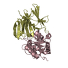
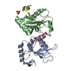
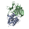
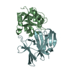
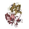
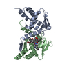
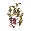
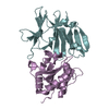
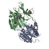
- PDB-4eqa: Crystal structure of PA1844 in complex with PA1845 from Pseudomon... -
+ Open data

- Basic information
| Entry | Database: PDB / ID: 4eqa | ||||||
|---|---|---|---|---|---|---|---|
| Title | Crystal structure of PA1844 in complex with PA1845 from Pseudomonas aeruginosa PAO1 | ||||||
 Components Components | (Putative uncharacterized protein) x 2 | ||||||
 Keywords Keywords | UNKNOWN FUNCTION / TYPE VI SECRETION / T6S / ANTITOXIN-TOXIN complex | ||||||
| Function / homology |  Function and homology information Function and homology informationgamma-D-glutamyl-meso-diaminopimelate peptidase / amidase activity / host cell membrane / extracellular region Similarity search - Function | ||||||
| Biological species |   Pseudomonas aeruginosa (bacteria) Pseudomonas aeruginosa (bacteria) | ||||||
| Method |  X-RAY DIFFRACTION / SYNCHROTRON / MOLECULAR REPLACEMENT / Resolution: 1.6 Å X-RAY DIFFRACTION / SYNCHROTRON / MOLECULAR REPLACEMENT / Resolution: 1.6 Å | ||||||
 Authors Authors | Shang, G. / Li, N. / Zhang, J. / Lu, D. / Yu, Q. / Zhao, Y. / Liu, X. / Xu, S. / Gu, L. | ||||||
 Citation Citation | Journal: Biochem.J. / Year: 2012 Title: Structural insight into how Pseudomonas aeruginosa peptidoglycanhydrolase Tse1 and its immunity protein Tsi1 function. Authors: Shang, G. / Liu, X. / Lu, D. / Zhang, J. / Li, N. / Zhu, C. / Liu, S. / Yu, Q. / Zhao, Y. / Zhang, H. / Hu, J. / Cang, H. / Xu, S. / Gu, L. | ||||||
| History |
|
- Structure visualization
| Structure viewer | Molecule: MolmilJmol/JSmol |
|---|
- Downloads & links
-Download
| PDBx/mmCIF format | 4eqa.cif.gz | 268.3 KB | Display | PDBx/mmCIF format |
|---|---|---|---|---|
| PDB format | pdb4eqa.ent.gz | 217.3 KB | Display | PDB format |
| PDBx/mmJSON format | 4eqa.json.gz | Tree view | PDBx/mmJSON format | |
| Others |  Other downloads Other downloads |
-Validation report
| Arichive directory | https://data.pdbj.org/pub/pdb/validation_reports/eq/4eqaftp://data.pdbj.org/pub/pdb/validation_reports/eq/4eqa | HTTPS FTP |
|---|
-Related structure data
| Related structure data |  4eq8SC S: Starting model for refinement C: citing same article ( |
|---|---|
| Similar structure data |
-Links
 PDBj
PDBj- Assembly
| Deposited unit | 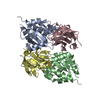
| ||||||||
|---|---|---|---|---|---|---|---|---|---|
| 1 | 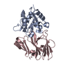
| ||||||||
| 2 | 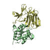
| ||||||||
| Unit cell |
|
-Components
| #1: Protein | Mass: 15640.782 Da / Num. of mol.: 2 / Fragment: The trunction, UNP residues 6-148 Source method: isolated from a genetically manipulated source Source: (gene. exp.) Pseudomonas aeruginosa (bacteria) / Strain: PAO1 / Gene: PA1844 / Plasmid: pET28b / Production host: #2: Protein | Mass: 16855.641 Da / Num. of mol.: 2 / Fragment: The trunction, UNP residues 20-172 Source method: isolated from a genetically manipulated source Source: (gene. exp.) Pseudomonas aeruginosa (bacteria) / Strain: PAO1 / Gene: PA1845 / Plasmid: pET21b / Production host: #3: Water | ChemComp-HOH / |  Mass: 18.015 Da / Num. of mol.: 779 / Source method: isolated from a natural source / Formula: H2O Mass: 18.015 Da / Num. of mol.: 779 / Source method: isolated from a natural source / Formula: H2OHas protein modification | Y | |
|---|
-Experimental details
-Experiment
| Experiment | Method: X-RAY DIFFRACTION / Number of used crystals: 1 |
|---|
- Sample preparation
| Crystal | Density Matthews: 2.06 Å3/Da / Density % sol: 40.32 % |
|---|---|
| Crystal grow | Temperature: 293 K / Method: vapor diffusion, hanging drop / pH: 6.5 Details: 20% PEG 3350, 0.1M Bis-Tris pH 6.5, VAPOR DIFFUSION, HANGING DROP, temperature 293K |
-Data collection
| Diffraction | Mean temperature: 100 K |
|---|---|
| Diffraction source | Source: SYNCHROTRON / Site: SSRF  / Beamline: BL17U / Wavelength: 0.9792 Å / Beamline: BL17U / Wavelength: 0.9792 Å |
| Detector | Type: ADSC QUANTUM 315r / Detector: CCD / Date: Nov 8, 2011 |
| Radiation | Monochromator: SAGITALLY FOCUSED SI(111) / Protocol: SINGLE WAVELENGTH / Monochromatic (M) / Laue (L): M / Scattering type: x-ray |
| Radiation wavelength | Wavelength: 0.9792 Å / Relative weight: 1 |
| Reflection | Resolution: 1.6→50 Å / Num. all: 68954 / Num. obs: 68954 / % possible obs: 99.6 % / Observed criterion σ(F): 0 / Observed criterion σ(I): 0 / Redundancy: 3.7 % / Biso Wilson estimate: 23.2 Å2 / Rmerge(I) obs: 0.108 / Rsym value: 0.108 / Net I/σ(I): 18.3 |
| Reflection shell | Resolution: 1.6→1.66 Å / Redundancy: 3.6 % / Rmerge(I) obs: 0.364 / Mean I/σ(I) obs: 7.9 / Num. unique all: 6876 / Rsym value: 0.364 / % possible all: 100 |
- Processing
| Software |
| ||||||||||||||||||||||||||||||||||||||||||||||||||||||||||||||||||||||||||||||||||||||||||
|---|---|---|---|---|---|---|---|---|---|---|---|---|---|---|---|---|---|---|---|---|---|---|---|---|---|---|---|---|---|---|---|---|---|---|---|---|---|---|---|---|---|---|---|---|---|---|---|---|---|---|---|---|---|---|---|---|---|---|---|---|---|---|---|---|---|---|---|---|---|---|---|---|---|---|---|---|---|---|---|---|---|---|---|---|---|---|---|---|---|---|---|
| Refinement | Method to determine structure: MOLECULAR REPLACEMENT Starting model: 4EQ8 Resolution: 1.6→27.22 Å / SU ML: 0.16 / σ(F): 0 / σ(I): 0 / Phase error: 21.13 / Stereochemistry target values: ML
| ||||||||||||||||||||||||||||||||||||||||||||||||||||||||||||||||||||||||||||||||||||||||||
| Solvent computation | Shrinkage radii: 0.9 Å / VDW probe radii: 1.11 Å / Solvent model: FLAT BULK SOLVENT MODEL / Bsol: 45.564 Å2 / ksol: 0.354 e/Å3 | ||||||||||||||||||||||||||||||||||||||||||||||||||||||||||||||||||||||||||||||||||||||||||
| Displacement parameters |
| ||||||||||||||||||||||||||||||||||||||||||||||||||||||||||||||||||||||||||||||||||||||||||
| Refinement step | Cycle: LAST / Resolution: 1.6→27.22 Å
| ||||||||||||||||||||||||||||||||||||||||||||||||||||||||||||||||||||||||||||||||||||||||||
| Refine LS restraints |
| ||||||||||||||||||||||||||||||||||||||||||||||||||||||||||||||||||||||||||||||||||||||||||
| LS refinement shell | Refine-ID: X-RAY DIFFRACTION / Total num. of bins used: 14
| ||||||||||||||||||||||||||||||||||||||||||||||||||||||||||||||||||||||||||||||||||||||||||
| Refinement TLS params. | Method: refined / Origin x: -11.2997 Å / Origin y: 24.3289 Å / Origin z: -15.2746 Å
| ||||||||||||||||||||||||||||||||||||||||||||||||||||||||||||||||||||||||||||||||||||||||||
| Refinement TLS group | Selection details: ALL |
