 Movie
Movie Controller


+ Open data

- Basic information
| Entry | Database: PDB / ID: 3p19 | ||||||
|---|---|---|---|---|---|---|---|
| Title | Improved NADPH-dependent Blue Fluorescent Protein | ||||||
 Components Components | Putative blue fluorescent protein | ||||||
 Keywords Keywords | OXIDOREDUCTASE / Rossmann-fold / Blue fluorescent protein | ||||||
| Function / homology |  Function and homology information Function and homology informationoxidoreductase activity, acting on the CH-OH group of donors, NAD or NADP as acceptor / nucleotide binding Similarity search - Function | ||||||
| Biological species |  Vibrio vulnificus (bacteria) Vibrio vulnificus (bacteria) | ||||||
| Method |  X-RAY DIFFRACTION / SYNCHROTRON / MOLECULAR REPLACEMENT / Resolution: 2.05 Å X-RAY DIFFRACTION / SYNCHROTRON / MOLECULAR REPLACEMENT / Resolution: 2.05 Å | ||||||
 Authors Authors | Kao, T.H. / Chen, Y. / Pai, C.H. / Wang, A.H.J. | ||||||
 Citation Citation | Journal: J.Struct.Biol. / Year: 2011 Title: Structure of a NADPH-dependent blue fluorescent protein revealed the unique role of Gly176 on the fluorescence enhancement. Authors: Kao, T.H. / Chen, Y. / Pai, C.H. / Chang, M.C. / Wang, A.H.J. | ||||||
| History |
|
- Structure visualization
| Structure viewer | Molecule: MolmilJmol/JSmol |
|---|
- Downloads & links
-Download
| PDBx/mmCIF format | 3p19.cif.gz | 197.5 KB | Display | PDBx/mmCIF format |
|---|---|---|---|---|
| PDB format | pdb3p19.ent.gz | 158.1 KB | Display | PDB format |
| PDBx/mmJSON format | 3p19.json.gz | Tree view | PDBx/mmJSON format | |
| Others |  Other downloads Other downloads |
-Validation report
| Arichive directory | https://data.pdbj.org/pub/pdb/validation_reports/p1/3p19ftp://data.pdbj.org/pub/pdb/validation_reports/p1/3p19 | HTTPS FTP |
|---|
-Related structure data
| Related structure data | 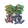 2japS S: Starting model for refinement |
|---|---|
| Similar structure data |
-Links
 PDBj
PDBj


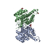
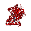
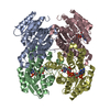
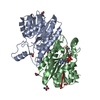
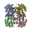
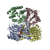
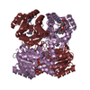
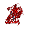
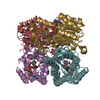
- Assembly
| Deposited unit | 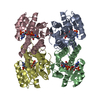
| ||||||||
|---|---|---|---|---|---|---|---|---|---|
| 1 |
| ||||||||
| 2 | 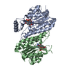
| ||||||||
| 3 | 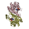
| ||||||||
| Unit cell |
|
-Components
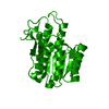
| #1: Protein | Mass: 28891.227 Da / Num. of mol.: 4 Source method: isolated from a genetically manipulated source Source: (gene. exp.) Vibrio vulnificus (bacteria) / Plasmid: pET21 / Production host: #2: Chemical | ChemComp-NDP /   Mass: 745.421 Da / Num. of mol.: 4 / Source method: obtained synthetically / Formula: C21H30N7O17P3 Mass: 745.421 Da / Num. of mol.: 4 / Source method: obtained synthetically / Formula: C21H30N7O17P3#3: Water | ChemComp-HOH / |  Mass: 18.015 Da / Num. of mol.: 543 / Source method: isolated from a natural source / Formula: H2O Mass: 18.015 Da / Num. of mol.: 543 / Source method: isolated from a natural source / Formula: H2OSequence details | A SEQUENCE DATABASE REFERENCE FOR THIS PROTEIN DOES NOT CURRENTLY EXIST. | |
|---|
-Experimental details
-Experiment
| Experiment | Method: X-RAY DIFFRACTION / Number of used crystals: 1 |
|---|
- Sample preparation
| Crystal | Density Matthews: 2.36 Å3/Da / Density % sol: 47.8 % |
|---|---|
| Crystal grow | Temperature: 277 K / Method: vapor diffusion, sitting drop / pH: 7.5 Details: 0.2M sodium citrate dihydrate, 0.1M HEPES-Na, 20% iso-propanol, pH 7.5, VAPOR DIFFUSION, SITTING DROP, temperature 277K |
-Data collection
| Diffraction | Mean temperature: 100 K |
|---|---|
| Diffraction source | Source: SYNCHROTRON / Site: NSRRC  / Beamline: BL13B1 / Wavelength: 1 Å / Beamline: BL13B1 / Wavelength: 1 Å |
| Detector | Type: ADSC QUANTUM 315 / Detector: CCD / Date: Aug 28, 2008 Details: Vertically Collimating Premirror, LN2-Cooled Fixed-Exit Double Crystal Si(111) Monochromator, Toroidal Focusing Mirror |
| Radiation | Monochromator: LN2-Cooled, Fixed-Exit Double Crystal Monochromator Protocol: SINGLE WAVELENGTH / Monochromatic (M) / Laue (L): M / Scattering type: x-ray |
| Radiation wavelength | Wavelength: 1 Å / Relative weight: 1 |
| Reflection | Resolution: 2.05→30 Å / Num. all: 66791 / Num. obs: 66383 / % possible obs: 99.4 % / Observed criterion σ(F): 0 / Observed criterion σ(I): 1 / Redundancy: 6.8 % / Rmerge(I) obs: 0.074 |
| Reflection shell | Resolution: 2.05→2.12 Å / Redundancy: 4.7 % / Rmerge(I) obs: 0.472 / Mean I/σ(I) obs: 2.293 / Num. unique all: 6413 / % possible all: 96.1 |
- Processing
| Software |
| |||||||||||||||||||||||||||||||||||||||||||||||||||||||||||||||||
|---|---|---|---|---|---|---|---|---|---|---|---|---|---|---|---|---|---|---|---|---|---|---|---|---|---|---|---|---|---|---|---|---|---|---|---|---|---|---|---|---|---|---|---|---|---|---|---|---|---|---|---|---|---|---|---|---|---|---|---|---|---|---|---|---|---|---|
| Refinement | Method to determine structure: MOLECULAR REPLACEMENT Starting model: 2JAP Resolution: 2.05→24.32 Å / Cor.coef. Fo:Fc: 0.963 / Cor.coef. Fo:Fc free: 0.938 / Occupancy max: 1 / Occupancy min: 1 / SU B: 5.055 / SU ML: 0.135 / Cross valid method: THROUGHOUT / σ(F): 0 / ESU R Free: 0.174 / Stereochemistry target values: MAXIMUM LIKELIHOOD Details: HYDROGENS HAVE BEEN ADDED IN THE RIDING POSITIONS U VALUES: REFINED INDIVIDUALLY
| |||||||||||||||||||||||||||||||||||||||||||||||||||||||||||||||||
| Solvent computation | Ion probe radii: 0.8 Å / Shrinkage radii: 0.8 Å / VDW probe radii: 1.4 Å / Solvent model: MASK | |||||||||||||||||||||||||||||||||||||||||||||||||||||||||||||||||
| Displacement parameters | Biso max: 80.86 Å2 / Biso mean: 35.538 Å2 / Biso min: 16.47 Å2
| |||||||||||||||||||||||||||||||||||||||||||||||||||||||||||||||||
| Refinement step | Cycle: LAST / Resolution: 2.05→24.32 Å
| |||||||||||||||||||||||||||||||||||||||||||||||||||||||||||||||||
| Refine LS restraints |
| |||||||||||||||||||||||||||||||||||||||||||||||||||||||||||||||||
| LS refinement shell | Resolution: 2.05→2.103 Å / Total num. of bins used: 20
|
