 Movie
Movie Controller


+ Open data

- Basic information
| Entry | Database: PDB / ID: 1gh6 | ||||||
|---|---|---|---|---|---|---|---|
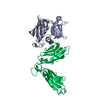
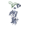
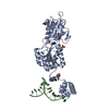
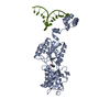
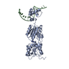
| Title | RETINOBLASTOMA POCKET COMPLEXED WITH SV40 LARGE T ANTIGEN | ||||||
 Components Components |
| ||||||
 Keywords Keywords | ANTITUMOR PROTEIN / TUMOR SUPPRESSOR / ONCOPROTEIN | ||||||
| Function / homology |  Function and homology information Function and homology informationDefective translocation of RB1 mutants to the nucleus / Rb-E2F complex / regulation of lipid kinase activity / positive regulation of collagen fibril organization / maintenance of mitotic sister chromatid cohesion / negative regulation of myofibroblast differentiation / cell morphogenesis involved in neuron differentiation / Positive Regulation of CDH1 Gene Transcription / sister chromatid biorientation / positive regulation of transcription regulatory region DNA binding ...Defective translocation of RB1 mutants to the nucleus / Rb-E2F complex / regulation of lipid kinase activity / positive regulation of collagen fibril organization / maintenance of mitotic sister chromatid cohesion / negative regulation of myofibroblast differentiation / cell morphogenesis involved in neuron differentiation / Positive Regulation of CDH1 Gene Transcription / sister chromatid biorientation / positive regulation of transcription regulatory region DNA binding / symbiont-mediated suppression of host JAK-STAT cascade via inhibition of JAK1 activity / positive regulation of extracellular matrix organization / Aberrant regulation of mitotic exit in cancer due to RB1 defects / negative regulation of hepatocyte apoptotic process / Inhibition of replication initiation of damaged DNA by RB1/E2F1 / protein localization to chromosome, centromeric region / importin-alpha family protein binding / myoblast differentiation / positive regulation of mitotic metaphase/anaphase transition / bidirectional double-stranded viral DNA replication / viral DNA genome replication / Replication of the SARS-CoV-1 genome / aortic valve morphogenesis / negative regulation of cold-induced thermogenesis / SWI/SNF complex / Formation of Senescence-Associated Heterochromatin Foci (SAHF) / negative regulation of G1/S transition of mitotic cell cycle / Phosphorylation of proteins involved in G1/S transition by active Cyclin E:Cdk2 complexes / RUNX2 regulates osteoblast differentiation / symbiont-mediated perturbation of host cell cycle G1/S transition checkpoint / Defective binding of RB1 mutants to E2F1,(E2F2, E2F3) / negative regulation of cell cycle / 3'-5' DNA helicase activity / DNA 3'-5' helicase / chondrocyte differentiation / DNA replication origin binding / negative regulation of apoptotic signaling pathway / chromosome organization / Cyclin E associated events during G1/S transition / negative regulation of protein kinase activity / Cyclin A:Cdk2-associated events at S phase entry / Nuclear events stimulated by ALK signaling in cancer / regulation of mitotic cell cycle / Condensation of Prophase Chromosomes / RNA polymerase II transcription regulatory region sequence-specific DNA binding / phosphoprotein binding / negative regulation of cell growth / negative regulation of inflammatory response / PML body / Oncogene Induced Senescence / APC/C:Cdh1 mediated degradation of Cdc20 and other APC/C:Cdh1 targeted proteins in late mitosis/early G1 / spindle / kinase binding / neuron projection development / cellular response to insulin stimulus / disordered domain specific binding / Cyclin D associated events in G1 / transcription corepressor activity / single-stranded DNA binding / heterochromatin formation / MLL4 and MLL3 complexes regulate expression of PPARG target genes in adipogenesis and hepatic steatosis / double-stranded DNA binding / Replication of the SARS-CoV-2 genome / DNA-binding transcription factor binding / spermatogenesis / molecular adaptor activity / symbiont-mediated perturbation of host ubiquitin-like protein modification / RNA polymerase II-specific DNA-binding transcription factor binding / Ras protein signal transduction / cell differentiation / DNA replication / regulation of cell cycle / symbiont-mediated suppression of host innate immune response / symbiont-mediated suppression of host type I interferon-mediated signaling pathway / chromatin remodeling / negative regulation of gene expression / negative regulation of DNA-templated transcription / apoptotic process / ubiquitin protein ligase binding / regulation of DNA-templated transcription / chromatin / host cell nucleus / negative regulation of transcription by RNA polymerase II / DNA-templated transcription / ATP hydrolysis activity / zinc ion binding / nucleoplasm / ATP binding / identical protein binding / nucleus / cytosol / cytoplasm Similarity search - Function | ||||||
| Biological species |  Simian virus 40 Simian virus 40 Homo sapiens (human) Homo sapiens (human) | ||||||
| Method |  X-RAY DIFFRACTION / MOLECULAR REPLACEMENT / Resolution: 3.2 Å X-RAY DIFFRACTION / MOLECULAR REPLACEMENT / Resolution: 3.2 Å | ||||||
 Authors Authors | Kim, H.Y. / Cho, Y. | ||||||
 Citation Citation | Journal: EMBO J. / Year: 2001 Title: Structural basis for the inactivation of retinoblastoma tumor suppressor by SV40 large T antigen. Authors: Kim, H.Y. / Ahn, B.Y. / Cho, Y. | ||||||
| History |
|
- Structure visualization
| Structure viewer | Molecule: MolmilJmol/JSmol |
|---|
- Downloads & links
-Download
| PDBx/mmCIF format | 1gh6.cif.gz | 102 KB | Display | PDBx/mmCIF format |
|---|---|---|---|---|
| PDB format | pdb1gh6.ent.gz | 78 KB | Display | PDB format |
| PDBx/mmJSON format | 1gh6.json.gz | Tree view | PDBx/mmJSON format | |
| Others |  Other downloads Other downloads |
-Validation report
| Arichive directory | https://data.pdbj.org/pub/pdb/validation_reports/gh/1gh6ftp://data.pdbj.org/pub/pdb/validation_reports/gh/1gh6 | HTTPS FTP |
|---|
-Related structure data
| Related structure data | 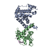 1guxS S: Starting model for refinement |
|---|---|
| Similar structure data |


-Links
 PDBj
PDBj


- Assembly
| Deposited unit | 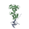
| ||||||||
|---|---|---|---|---|---|---|---|---|---|
| 1 |
| ||||||||
| Unit cell |
|
-Components
| #1: Protein | Mass: 13436.985 Da / Num. of mol.: 1 Source method: isolated from a genetically manipulated source Source: (gene. exp.) Simian virus 40 / Genus: Polyomavirus / Plasmid: PET15 / Species (production host): Escherichia coli / Production host:  References: UniProt: P03070, Hydrolases; Acting on acid anhydrides; Acting on acid anhydrides to facilitate cellular and subcellular movement |
|---|---|
| #2: Protein | Mass: 39026.504 Da / Num. of mol.: 1 / Fragment: UNP residues 379-577 and 645-772 Source method: isolated from a genetically manipulated source Source: (gene. exp.) Homo sapiens / Production host: |
-Experimental details
-Experiment
| Experiment | Method: X-RAY DIFFRACTION / Number of used crystals: 1 |
|---|
- Sample preparation
| Crystal | Density Matthews: 3.7 Å3/Da / Density % sol: 66.79 % | |||||||||||||||||||||||||||||||||||||||||||||||||||||||||||||||
|---|---|---|---|---|---|---|---|---|---|---|---|---|---|---|---|---|---|---|---|---|---|---|---|---|---|---|---|---|---|---|---|---|---|---|---|---|---|---|---|---|---|---|---|---|---|---|---|---|---|---|---|---|---|---|---|---|---|---|---|---|---|---|---|---|
| Crystal grow | pH: 7.2 / Details: pH 7.2 | |||||||||||||||||||||||||||||||||||||||||||||||||||||||||||||||
| Crystal grow | *PLUS Temperature: 4 ℃ / pH: 7.5 / Method: vapor diffusion, hanging drop | |||||||||||||||||||||||||||||||||||||||||||||||||||||||||||||||
| Components of the solutions | *PLUS
|
-Data collection
| Diffraction | Mean temperature: 200 K |
|---|---|
| Diffraction source | Source: ROTATING ANODE / Type: RIGAKU RU200 / Wavelength: 1.5418 |
| Detector | Type: MARRESEARCH / Detector: IMAGE PLATE / Date: Mar 1, 2000 / Details: MIRRORS |
| Radiation | Protocol: SINGLE WAVELENGTH / Monochromatic (M) / Laue (L): M / Scattering type: x-ray |
| Radiation wavelength | Wavelength: 1.5418 Å / Relative weight: 1 |
| Reflection | Resolution: 3.2→99 Å / Num. obs: 12666 / % possible obs: 99.5 % / Observed criterion σ(I): 1 / Redundancy: 4.2 % / Rmerge(I) obs: 0.084 / Rsym value: 0.106 / Net I/σ(I): 15.9 |
| Reflection shell | Resolution: 3.2→3.29 Å / Redundancy: 4 % / Rmerge(I) obs: 0.377 / Mean I/σ(I) obs: 2.8 / Rsym value: 0.369 / % possible all: 97.2 |
| Reflection | *PLUS Lowest resolution: 20 Å / Num. measured all: 50139 |
| Reflection shell | *PLUS % possible obs: 97.2 % / Rmerge(I) obs: 0.369 |
- Processing
| Software |
| ||||||||||||||||||||||||||||||||||||||||||||||||||||||||||||
|---|---|---|---|---|---|---|---|---|---|---|---|---|---|---|---|---|---|---|---|---|---|---|---|---|---|---|---|---|---|---|---|---|---|---|---|---|---|---|---|---|---|---|---|---|---|---|---|---|---|---|---|---|---|---|---|---|---|---|---|---|---|
| Refinement | Method to determine structure: MOLECULAR REPLACEMENT Starting model: 1GUX Resolution: 3.2→19.92 Å / Rfactor Rfree error: 0.013 / Cross valid method: THROUGHOUT / σ(F): 0
| ||||||||||||||||||||||||||||||||||||||||||||||||||||||||||||
| Displacement parameters | Biso mean: 44.2 Å2
| ||||||||||||||||||||||||||||||||||||||||||||||||||||||||||||
| Refinement step | Cycle: LAST / Resolution: 3.2→19.92 Å
| ||||||||||||||||||||||||||||||||||||||||||||||||||||||||||||
| Refine LS restraints |
| ||||||||||||||||||||||||||||||||||||||||||||||||||||||||||||
| LS refinement shell | Resolution: 3.2→3.4 Å / Rfactor Rfree error: 0.044 / Total num. of bins used: 6
| ||||||||||||||||||||||||||||||||||||||||||||||||||||||||||||
| Xplor file |
| ||||||||||||||||||||||||||||||||||||||||||||||||||||||||||||
| Software | *PLUS Name: CNS / Version: 1 / Classification: refinement | ||||||||||||||||||||||||||||||||||||||||||||||||||||||||||||
| Refinement | *PLUS Highest resolution: 3.2 Å / σ(F): 0 / % reflection Rfree: 5 % | ||||||||||||||||||||||||||||||||||||||||||||||||||||||||||||
| Solvent computation | *PLUS | ||||||||||||||||||||||||||||||||||||||||||||||||||||||||||||
| Displacement parameters | *PLUS Biso mean: 44.2 Å2 | ||||||||||||||||||||||||||||||||||||||||||||||||||||||||||||
| Refine LS restraints | *PLUS Type: c_angle_deg / Dev ideal: 1.44 | ||||||||||||||||||||||||||||||||||||||||||||||||||||||||||||
| LS refinement shell | *PLUS Rfactor Rfree: 0.445 / % reflection Rfree: 5.5 % / Rfactor Rwork: 0.377 |
