 Movie
Movie Controller

[English] 日本語
 Yorodumi
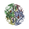
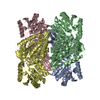
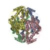
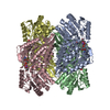
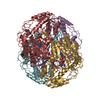
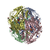
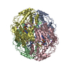
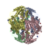
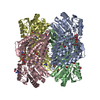
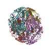
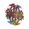
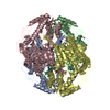
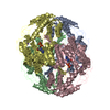
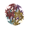
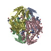
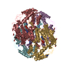
- PDB-1o01: Human mitochondrial aldehyde dehydrogenase complexed with crotona... -
+ Open data

- Basic information
| Entry | Database: PDB / ID: 1o01 | ||||||
|---|---|---|---|---|---|---|---|
| Title | Human mitochondrial aldehyde dehydrogenase complexed with crotonaldehyde, NAD(H) and Mg2+ | ||||||
 Components Components | Aldehyde dehydrogenase | ||||||
 Keywords Keywords | OXIDOREDUCTASE / ALDH / NAD / NADH / isomerization | ||||||
| Function / homology |  Function and homology information Function and homology informationMetabolism of serotonin / nitroglycerin metabolic process / regulation of dopamine biosynthetic process / regulation of serotonin biosynthetic process / phenylacetaldehyde dehydrogenase (NAD+) activity / ethanol metabolic process / aldehyde catabolic process / alcohol metabolic process / ethanol catabolic process / Ethanol oxidation ...Metabolism of serotonin / nitroglycerin metabolic process / regulation of dopamine biosynthetic process / regulation of serotonin biosynthetic process / phenylacetaldehyde dehydrogenase (NAD+) activity / ethanol metabolic process / aldehyde catabolic process / alcohol metabolic process / ethanol catabolic process / Ethanol oxidation / aldehyde dehydrogenase [NAD(P)+] activity / aldehyde dehydrogenase (NAD+) / carboxylesterase activity / cellular detoxification of aldehyde / aldehyde dehydrogenase (NAD+) activity / Smooth Muscle Contraction / Mitochondrial protein degradation / NAD binding / carbohydrate metabolic process / electron transfer activity / mitochondrial matrix / mitochondrion / extracellular exosome Similarity search - Function | ||||||
| Biological species |  Homo sapiens (human) Homo sapiens (human) | ||||||
| Method |  X-RAY DIFFRACTION / SYNCHROTRON / MOLECULAR REPLACEMENT / Resolution: 2.15 Å X-RAY DIFFRACTION / SYNCHROTRON / MOLECULAR REPLACEMENT / Resolution: 2.15 Å | ||||||
 Authors Authors | Perez-Miller, S.J. / Hurley, T.D. | ||||||
 Citation Citation | Journal: Biochemistry / Year: 2003 Title: Coenzyme isomerization is integral to catalysis in aldehyde dehydrogenase Authors: Perez-Miller, S.J. / Hurley, T.D. | ||||||
| History |
|
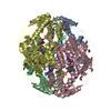
- Structure visualization
| Structure viewer | Molecule: MolmilJmol/JSmol |
|---|
- Downloads & links
-Download
| PDBx/mmCIF format | 1o01.cif.gz | 833.8 KB | Display | PDBx/mmCIF format |
|---|---|---|---|---|
| PDB format | pdb1o01.ent.gz | 684.6 KB | Display | PDB format |
| PDBx/mmJSON format | 1o01.json.gz | Tree view | PDBx/mmJSON format | |
| Others |  Other downloads Other downloads |
-Validation report
| Arichive directory | https://data.pdbj.org/pub/pdb/validation_reports/o0/1o01ftp://data.pdbj.org/pub/pdb/validation_reports/o0/1o01 | HTTPS FTP |
|---|
-Related structure data
| Related structure data |  1nzwC  1nzxC 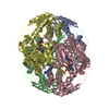 1nzzC  1o00C  1o02C  1o04C 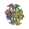 1cw3S S: Starting model for refinement C: citing same article ( |
|---|---|
| Similar structure data |

-Links
 PDBj
PDBj


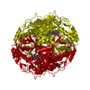
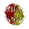
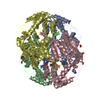
- Assembly
| Deposited unit | 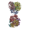
| ||||||||
|---|---|---|---|---|---|---|---|---|---|
| 1 | 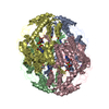
| ||||||||
| 2 | 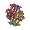
| ||||||||
| Unit cell |
|
-Components
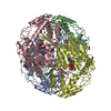
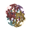
-Protein , 1 types, 8 molecules ABCDEFGH
| #1: Protein | Mass: 54499.629 Da / Num. of mol.: 8 Fragment: Complete mature sequence (does not contain mitochondrial leader sequence). Source method: isolated from a genetically manipulated source Source: (gene. exp.) Homo sapiens (human) / Gene: ALDH2 OR ALDM / Plasmid: PET7-7 / Species (production host): Escherichia coli / Production host:  |
|---|
-Non-polymers , 7 types, 3711 molecules 

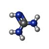


| #2: Chemical | ChemComp-MG /  Mass: 24.305 Da / Num. of mol.: 8 / Source method: obtained synthetically / Formula: Mg Mass: 24.305 Da / Num. of mol.: 8 / Source method: obtained synthetically / Formula: Mg#3: Chemical | ChemComp-NA /  Mass: 22.990 Da / Num. of mol.: 8 / Source method: obtained synthetically / Formula: Na Mass: 22.990 Da / Num. of mol.: 8 / Source method: obtained synthetically / Formula: Na#4: Chemical | ChemComp-GAI /  Mass: 59.070 Da / Num. of mol.: 8 / Source method: obtained synthetically / Formula: CH5N3 Mass: 59.070 Da / Num. of mol.: 8 / Source method: obtained synthetically / Formula: CH5N3#5: Chemical | ChemComp-NAD /  Mass: 663.425 Da / Num. of mol.: 8 / Source method: obtained synthetically / Formula: C21H27N7O14P2 / Comment: NAD*YM Mass: 663.425 Da / Num. of mol.: 8 / Source method: obtained synthetically / Formula: C21H27N7O14P2 / Comment: NAD*YM#6: Chemical | ChemComp-CRD / (  Mass: 70.090 Da / Num. of mol.: 6 / Source method: obtained synthetically / Formula: C4H6O Mass: 70.090 Da / Num. of mol.: 6 / Source method: obtained synthetically / Formula: C4H6O#7: Chemical | ChemComp-EDO /  Mass: 62.068 Da / Num. of mol.: 8 / Source method: obtained synthetically / Formula: C2H6O2 Mass: 62.068 Da / Num. of mol.: 8 / Source method: obtained synthetically / Formula: C2H6O2#8: Water | ChemComp-HOH / | Mass: 18.015 Da / Num. of mol.: 3665 / Source method: isolated from a natural source / Formula: H2O |
|---|
-Experimental details
-Experiment
| Experiment | Method: X-RAY DIFFRACTION / Number of used crystals: 1 |
|---|
- Sample preparation
| Crystal | Density Matthews: 1.95 Å3/Da / Density % sol: 36.57 % | |||||||||||||||||||||||||||||||||||||||||||||||||
|---|---|---|---|---|---|---|---|---|---|---|---|---|---|---|---|---|---|---|---|---|---|---|---|---|---|---|---|---|---|---|---|---|---|---|---|---|---|---|---|---|---|---|---|---|---|---|---|---|---|---|
| Crystal grow | Temperature: 293 K / Method: vapor diffusion, sitting drop / pH: 6.4 Details: ACES, PEG 6000, Guanidine HCl, MgCl2, DTT, NAD+. Crystal soaked with crotonaldehyde prior to data collection., pH 6.4, VAPOR DIFFUSION, SITTING DROP, temperature 293K | |||||||||||||||||||||||||||||||||||||||||||||||||
| Crystal grow | *PLUS Temperature: 20 ℃ / Method: vapor diffusion, sitting drop / PH range low: 6.6 / PH range high: 6.2 | |||||||||||||||||||||||||||||||||||||||||||||||||
| Components of the solutions | *PLUS
|
-Data collection
| Diffraction | Mean temperature: 100 K |
|---|---|
| Diffraction source | Source: SYNCHROTRON / Site: NSLS  / Beamline: X12C / Wavelength: 0.9786 Å / Beamline: X12C / Wavelength: 0.9786 Å |
| Detector | Type: BRANDEIS - B4 / Detector: CCD / Date: Aug 29, 1999 |
| Radiation | Monochromator: double crystal / Protocol: SINGLE WAVELENGTH / Monochromatic (M) / Laue (L): M / Scattering type: x-ray |
| Radiation wavelength | Wavelength: 0.9786 Å / Relative weight: 1 |
| Reflection | Resolution: 2.15→35 Å / Num. all: 207332 / Num. obs: 188672 / % possible obs: 91 % / Observed criterion σ(F): 0 / Observed criterion σ(I): 0 / Redundancy: 3.6 % / Biso Wilson estimate: 16 Å2 / Rmerge(I) obs: 0.059 / Net I/σ(I): 19.5 |
| Reflection shell | Resolution: 2.15→2.23 Å / Rmerge(I) obs: 0.136 / Mean I/σ(I) obs: 7.2 / Num. unique all: 14801 / % possible all: 72 |
| Reflection | *PLUS Lowest resolution: 45 Å / % possible obs: 90.7 % / Num. measured all: 688308 |
| Reflection shell | *PLUS % possible obs: 71.9 % |
- Processing
| Software |
| ||||||||||||||||||||||||||||||||||||
|---|---|---|---|---|---|---|---|---|---|---|---|---|---|---|---|---|---|---|---|---|---|---|---|---|---|---|---|---|---|---|---|---|---|---|---|---|---|
| Refinement | Method to determine structure: MOLECULAR REPLACEMENT Starting model: 1CW3 Resolution: 2.15→34.84 Å / Rfactor Rfree error: 0.002 / Data cutoff high absF: 6380979.5 / Data cutoff high rms absF: 6380979.5 / Data cutoff low absF: 0 / Isotropic thermal model: RESTRAINED / Cross valid method: THROUGHOUT / σ(F): 0 / σ(I): 0 / Stereochemistry target values: Engh & Huber / Details: maximum likelihood target using amplitudes
| ||||||||||||||||||||||||||||||||||||
| Solvent computation | Solvent model: FLAT MODEL / Bsol: 20.2129 Å2 / ksol: 0.310269 e/Å3 | ||||||||||||||||||||||||||||||||||||
| Displacement parameters | Biso mean: 19.5 Å2
| ||||||||||||||||||||||||||||||||||||
| Refine analyze |
| ||||||||||||||||||||||||||||||||||||
| Refinement step | Cycle: LAST / Resolution: 2.15→34.84 Å
| ||||||||||||||||||||||||||||||||||||
| Refine LS restraints |
| ||||||||||||||||||||||||||||||||||||
| Refine LS restraints NCS | NCS model details: RESTRAINED | ||||||||||||||||||||||||||||||||||||
| LS refinement shell | Resolution: 2.15→2.23 Å / Rfactor Rfree error: 0.01 / Total num. of bins used: 10
| ||||||||||||||||||||||||||||||||||||
| Xplor file |
| ||||||||||||||||||||||||||||||||||||
| Refinement | *PLUS Lowest resolution: 45 Å / % reflection Rfree: 5 % / Rfactor Rfree: 0.207 / Rfactor Rwork: 0.17 | ||||||||||||||||||||||||||||||||||||
| Solvent computation | *PLUS | ||||||||||||||||||||||||||||||||||||
| Displacement parameters | *PLUS | ||||||||||||||||||||||||||||||||||||
| Refine LS restraints | *PLUS
|
