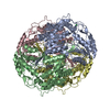
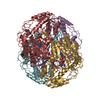
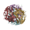
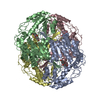
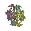
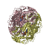
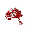
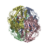
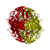
Entry Database : PDB / ID : 5l2mTitle Structure of ALDH1A1 in complex with BUC11 Retinal dehydrogenase 1 Keywords / / Function / homology Function Domain/homology Component
/ / / / / / / / / / / / / / / / / / / / / / / / / / / / / / / / / / / / / / / / / / / / / / / / / / / / / / / / / / / / / / Biological species Homo sapiens (human)Method / / Resolution : 1.7 Å Authors Buchman, C.D. / Hurley, T.D. Funding support Organization Grant number Country National Institutes of Health/National Cancer Institute (NIH/NCI) R21 CA198409
Journal : J. Med. Chem. / Year : 2017Title : Inhibition of the Aldehyde Dehydrogenase 1/2 Family by Psoralen and Coumarin Derivatives.Authors : Buchman, C.D. / Hurley, T.D. History Deposition Aug 2, 2016 Deposition site / Processing site Revision 1.0 Mar 8, 2017 Provider / Type Revision 1.1 Apr 5, 2017 Group Revision 1.2 Sep 20, 2017 Group / Category / Item Revision 1.3 Dec 4, 2019 Group / Category / Item Revision 1.4 Nov 6, 2024 Group Data collection / Database references ... Data collection / Database references / Derived calculations / Structure summary Category chem_comp_atom / chem_comp_bond ... chem_comp_atom / chem_comp_bond / database_2 / pdbx_entry_details / pdbx_modification_feature / pdbx_struct_conn_angle / struct_conn Item _database_2.pdbx_DOI / _database_2.pdbx_database_accession ... _database_2.pdbx_DOI / _database_2.pdbx_database_accession / _pdbx_struct_conn_angle.ptnr1_auth_seq_id / _pdbx_struct_conn_angle.ptnr1_symmetry / _pdbx_struct_conn_angle.ptnr3_auth_seq_id / _pdbx_struct_conn_angle.ptnr3_symmetry / _pdbx_struct_conn_angle.value / _struct_conn.pdbx_dist_value / _struct_conn.ptnr2_auth_seq_id / _struct_conn.ptnr2_symmetry
Show all Show less
 Movie
Movie Controller


 Open data
 Basic information
 Components
 Keywords
 Function and homology information
 Homo sapiens (human)
 X-RAY DIFFRACTION /
  Authors
 United States, 1items
  Citation
 Structure visualization
 Downloads & links
 Other downloads
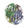


 PDBj
PDBj


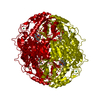
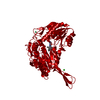
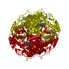
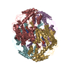
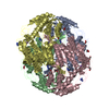
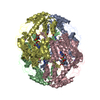
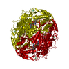
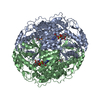
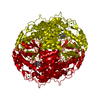
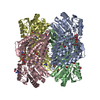
 Assembly
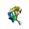

 Mass: 173.040 Da / Num. of mol.: 1 / Source method: obtained synthetically / Formula: Yb
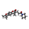
 Mass: 367.438 Da / Num. of mol.: 1 / Source method: obtained synthetically / Formula: C22H25NO4
 Mass: 35.453 Da / Num. of mol.: 1 / Source method: obtained synthetically / Formula: Cl
 Mass: 18.015 Da / Num. of mol.: 309 / Source method: isolated from a natural source / Formula: H2O
 Sample preparation
 Processing