 Movie
Movie Controller

[English] 日本語
 Yorodumi
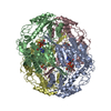
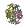
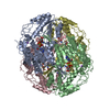
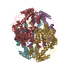
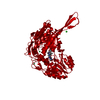
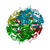
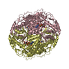
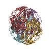
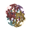
- PDB-1cw3: HUMAN MITOCHONDRIAL ALDEHYDE DEHYDROGENASE COMPLEXED WITH NAD+ AN... -
+ Open data

- Basic information
| Entry | Database: PDB / ID: 1cw3 | ||||||
|---|---|---|---|---|---|---|---|
| Title | HUMAN MITOCHONDRIAL ALDEHYDE DEHYDROGENASE COMPLEXED WITH NAD+ AND MN2+ | ||||||
 Components Components | MITOCHONDRIAL ALDEHYDE DEHYDROGENASE | ||||||
 Keywords Keywords | OXIDOREDUCTASE / DINUCLEOTIDE FOLD | ||||||
| Function / homology |  Function and homology information Function and homology informationMetabolism of serotonin / nitroglycerin metabolic process / regulation of dopamine biosynthetic process / regulation of serotonin biosynthetic process / phenylacetaldehyde dehydrogenase (NAD+) activity / ethanol metabolic process / aldehyde catabolic process / alcohol metabolic process / ethanol catabolic process / Ethanol oxidation ...Metabolism of serotonin / nitroglycerin metabolic process / regulation of dopamine biosynthetic process / regulation of serotonin biosynthetic process / phenylacetaldehyde dehydrogenase (NAD+) activity / ethanol metabolic process / aldehyde catabolic process / alcohol metabolic process / ethanol catabolic process / Ethanol oxidation / aldehyde dehydrogenase [NAD(P)+] activity / aldehyde dehydrogenase (NAD+) / carboxylesterase activity / cellular detoxification of aldehyde / aldehyde dehydrogenase (NAD+) activity / Smooth Muscle Contraction / Mitochondrial protein degradation / NAD binding / carbohydrate metabolic process / electron transfer activity / mitochondrial matrix / mitochondrion / extracellular exosome Similarity search - Function | ||||||
| Biological species |  Homo sapiens (human) Homo sapiens (human) | ||||||
| Method |  X-RAY DIFFRACTION / Resolution: 2.58 Å X-RAY DIFFRACTION / Resolution: 2.58 Å | ||||||
 Authors Authors | Ni, L. / Zhou, J. / Hurley, T.D. / Weiner, H. | ||||||
 Citation Citation | Journal: Protein Sci. / Year: 1999 Title: Human liver mitochondrial aldehyde dehydrogenase: three-dimensional structure and the restoration of solubility and activity of chimeric forms. Authors: Ni, L. / Zhou, J. / Hurley, T.D. / Weiner, H. #1: Journal: Structure / Year: 1997Title: Structure of mitochondrial aldehyde dehydrogenase: the genetic component of ethanol aversion. Authors: Steinmetz, C.G. / Xie, P. / Weiner, H. / Hurley, T.D. | ||||||
| History |
|
- Structure visualization
| Structure viewer | Molecule: MolmilJmol/JSmol |
|---|
- Downloads & links
-Download
| PDBx/mmCIF format | 1cw3.cif.gz | 767.6 KB | Display | PDBx/mmCIF format |
|---|---|---|---|---|
| PDB format | pdb1cw3.ent.gz | 636.9 KB | Display | PDB format |
| PDBx/mmJSON format | 1cw3.json.gz | Tree view | PDBx/mmJSON format | |
| Others |  Other downloads Other downloads |
-Validation report
| Arichive directory | https://data.pdbj.org/pub/pdb/validation_reports/cw/1cw3ftp://data.pdbj.org/pub/pdb/validation_reports/cw/1cw3 | HTTPS FTP |
|---|
-Related structure data
| Related structure data | |
|---|---|
| Similar structure data |

-Links
 PDBj
PDBj


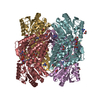
- Assembly
| Deposited unit | 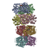
| ||||||||||
|---|---|---|---|---|---|---|---|---|---|---|---|
| 1 | 
| ||||||||||
| 2 | 
| ||||||||||
| Unit cell |
|
-Components
| #1: Protein | Mass: 53970.090 Da / Num. of mol.: 8 Fragment: COMPLETE MATURE SEQUENCE (DOES NOT INCLUDE MITOCHONDRIAL LEADER SEQUENCE) Source method: isolated from a genetically manipulated source Source: (gene. exp.) Homo sapiens (human) / Organ: LIVER / Plasmid: PET / Production host:  #2: Chemical | ChemComp-MN /   Mass: 54.938 Da / Num. of mol.: 8 / Source method: obtained synthetically / Formula: Mn Mass: 54.938 Da / Num. of mol.: 8 / Source method: obtained synthetically / Formula: Mn#3: Chemical | ChemComp-MG /   Mass: 24.305 Da / Num. of mol.: 8 / Source method: obtained synthetically / Formula: Mg Mass: 24.305 Da / Num. of mol.: 8 / Source method: obtained synthetically / Formula: Mg#4: Chemical | ChemComp-NAD /   Mass: 663.425 Da / Num. of mol.: 8 / Source method: obtained synthetically / Formula: C21H27N7O14P2 / Comment: NAD*YM Mass: 663.425 Da / Num. of mol.: 8 / Source method: obtained synthetically / Formula: C21H27N7O14P2 / Comment: NAD*YM#5: Water | ChemComp-HOH / |  Mass: 18.015 Da / Num. of mol.: 1087 / Source method: isolated from a natural source / Formula: H2O Mass: 18.015 Da / Num. of mol.: 1087 / Source method: isolated from a natural source / Formula: H2O |
|---|
-Experimental details
-Experiment
| Experiment | Method: X-RAY DIFFRACTION / Number of used crystals: 1 |
|---|
- Sample preparation
| Crystal | Density Matthews: 2.1 Å3/Da / Density % sol: 41.53 % | |||||||||||||||||||||||||||||||||||
|---|---|---|---|---|---|---|---|---|---|---|---|---|---|---|---|---|---|---|---|---|---|---|---|---|---|---|---|---|---|---|---|---|---|---|---|---|
| Crystal grow | Temperature: 293 K / Method: vapor diffusion, sitting drop / pH: 6.4 Details: 100 MM ACES, PH 6.4, 100 MM GUANIDINE-HCL, 2 MM NAD+, 4 MM DTT, 16% PEG 6000, 8 MG/ML ENZYME SOAKED WITH 8 MM MNCL2 FOR 2 DAYS, VAPOR DIFFUSION, SITTING DROP, temperature 293K | |||||||||||||||||||||||||||||||||||
| Crystal grow | *PLUS pH: 6.2 / Method: unknown | |||||||||||||||||||||||||||||||||||
| Components of the solutions | *PLUS
|
-Data collection
| Diffraction | Mean temperature: 113 K |
|---|---|
| Diffraction source | Source: ROTATING ANODE / Type: RIGAKU RU200 / Wavelength: 1.5418 |
| Detector | Type: RIGAKU RAXIS IIC / Detector: IMAGE PLATE / Date: Aug 8, 1998 |
| Radiation | Protocol: SINGLE WAVELENGTH / Monochromatic (M) / Laue (L): M / Scattering type: x-ray |
| Radiation wavelength | Wavelength: 1.5418 Å / Relative weight: 1 |
| Reflection | Resolution: 2.58→45 Å / Num. all: 112240 / Num. obs: 102859 / % possible obs: 91.6 % / Redundancy: 2.15 % / Biso Wilson estimate: 45 Å2 / Rmerge(I) obs: 0.072 / Net I/σ(I): 12.6 |
| Reflection shell | Resolution: 2.58→2.68 Å / Redundancy: 1.51 % / Rmerge(I) obs: 0.287 / % possible all: 64.3 |
| Reflection | *PLUS Lowest resolution: 45 Å / Num. measured all: 221417 |
| Reflection shell | *PLUS % possible obs: 64.3 % / Mean I/σ(I) obs: 3 |
- Processing
| Software |
| ||||||||||||||||||||||||||||||||||||||||||||||||||||||||||||
|---|---|---|---|---|---|---|---|---|---|---|---|---|---|---|---|---|---|---|---|---|---|---|---|---|---|---|---|---|---|---|---|---|---|---|---|---|---|---|---|---|---|---|---|---|---|---|---|---|---|---|---|---|---|---|---|---|---|---|---|---|---|
| Refinement | Resolution: 2.58→45 Å / Cross valid method: THROUGHOUT / σ(F): 0 / σ(I): 0 / Stereochemistry target values: ENGH & HUBER / Details: STANDARD X-PLOR WEIGHTS
| ||||||||||||||||||||||||||||||||||||||||||||||||||||||||||||
| Refinement step | Cycle: LAST / Resolution: 2.58→45 Å
| ||||||||||||||||||||||||||||||||||||||||||||||||||||||||||||
| Refine LS restraints |
| ||||||||||||||||||||||||||||||||||||||||||||||||||||||||||||
| Software | *PLUS Name: X-PLOR / Version: 3.851 / Classification: refinement | ||||||||||||||||||||||||||||||||||||||||||||||||||||||||||||
| Refinement | *PLUS Lowest resolution: 45 Å / σ(F): 0 / % reflection Rfree: 7 % / Rfactor obs: 0.175 | ||||||||||||||||||||||||||||||||||||||||||||||||||||||||||||
| Solvent computation | *PLUS | ||||||||||||||||||||||||||||||||||||||||||||||||||||||||||||
| Displacement parameters | *PLUS | ||||||||||||||||||||||||||||||||||||||||||||||||||||||||||||
| LS refinement shell | *PLUS Highest resolution: 2.58 Å / Lowest resolution: 2.68 Å |
