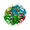
- PDB-6dum: ALDH1A1 N121S in complex with 6-{[(3-fluorophenyl)methyl]sulfanyl... -
+
Open data
ID or keywords:
Loading...
-
Basic information
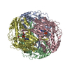
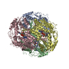
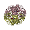
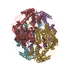
Entry
Database: PDB / ID: 6dum
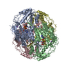
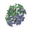
Title
ALDH1A1 N121S in complex with 6-{[(3-fluorophenyl)methyl]sulfanyl}-2-(oxetan-3-yl)-5-phenyl-2,5-dihydro-4H-pyrazolo[3,4-d]pyrimidin-4-one (compound 13g)
Retinaldehydrogenase1 / RalDH1 / ALDH-E1 / ALHDII / Aldehyde dehydrogenase family 1 member A1 / Aldehyde dehydrogenase / cytosolic
Mass: 54897.590 Da / Num. of mol.: 1 Source method: isolated from a genetically manipulated source Source: (gene. exp.) Homo sapiens (human) / Gene: ALDH1A1, ALDC, ALDH1, PUMB1 / Production host: Escherichia coli (E. coli) References: UniProt: V9HW83, UniProt: P00352*PLUS, Oxidoreductases; Acting on the aldehyde or oxo group of donors; With NAD+ or NADP+ as acceptor, retinal dehydrogenase
Resolution: 2→50 Å / Cor.coef. Fo:Fc: 0.964 / Cor.coef. Fo:Fc free: 0.934 / SU B: 15.824 / SU ML: 0.218 / Cross valid method: THROUGHOUT / σ(F): 0 / ESU R: 0.205 / ESU R Free: 0.183 Details: HYDROGENS HAVE BEEN ADDED IN THE RIDING POSITIONS U VALUES : WITH TLS ADDED
Rfactor
Num. reflection
% reflection
Selection details
Rfree
0.2486
1690
5.1 %
RANDOM
Rwork
0.1941
-
-
-
obs
0.1971
31637
97.15 %
-
Solvent computation
Ion probe radii: 0.8 Å / Shrinkage radii: 0.8 Å / VDW probe radii: 1.2 Å
In the structure databanks used in Yorodumi, some data are registered as the other names, "COVID-19 virus" and "2019-nCoV". Here are the details of the virus and the list of structure data.
Jan 31, 2019. EMDB accession codes are about to change! (news from PDBe EMDB page)
EMDB accession codes are about to change! (news from PDBe EMDB page)
The allocation of 4 digits for EMDB accession codes will soon come to an end. Whilst these codes will remain in use, new EMDB accession codes will include an additional digit and will expand incrementally as the available range of codes is exhausted. The current 4-digit format prefixed with “EMD-” (i.e. EMD-XXXX) will advance to a 5-digit format (i.e. EMD-XXXXX), and so on. It is currently estimated that the 4-digit codes will be depleted around Spring 2019, at which point the 5-digit format will come into force.
The EM Navigator/Yorodumi systems omit the EMD- prefix.
Related info.:Q: What is EMD? / ID/Accession-code notation in Yorodumi/EM Navigator
Yorodumi is a browser for structure data from EMDB, PDB, SASBDB, etc.
This page is also the successor to EM Navigator detail page, and also detail information page/front-end page for Omokage search.
The word "yorodu" (or yorozu) is an old Japanese word meaning "ten thousand". "mi" (miru) is to see.
Related info.:EMDB / PDB / SASBDB / Comparison of 3 databanks / Yorodumi Search / Aug 31, 2016. New EM Navigator & Yorodumi / Yorodumi Papers / Jmol/JSmol / Function and homology information / Changes in new EM Navigator and Yorodumi
 Movie
Movie Controller

 Yorodumi
 Open data
 Basic information
 Components
 Keywords
 Function and homology information
 Homo sapiens (human)
 X-RAY DIFFRACTION /
  Authors
 United States, 1items
  Citation
 Structure visualization
 Downloads & links
 Other downloads
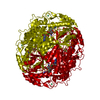


 PDBj
PDBj


 Assembly
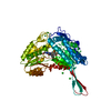


 Mass: 173.040 Da / Num. of mol.: 2 / Source method: obtained synthetically / Formula: Yb
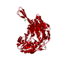
 Mass: 408.449 Da / Num. of mol.: 1 / Source method: obtained synthetically / Formula: C21H17FN4O2S
 Mass: 665.441 Da / Num. of mol.: 1 / Source method: obtained synthetically / Formula: C21H29N7O14P2
 Mass: 35.453 Da / Num. of mol.: 7 / Source method: obtained synthetically / Formula: Cl
 Sample preparation
 Processing