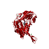
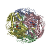
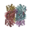
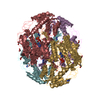
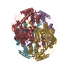
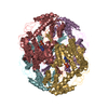
Entry Database : PDB / ID : 4wj9Title Structure of Human apo ALDH1A1 Retinal dehydrogenase 1 Keywords / Function / homology Function Domain/homology Component
/ / / / / / / / / / / / / / / / / / / / / / / / / / / / / / / / / / / / / / / / / / / / / / / / / / / / / / / / / / / / / / Biological species Homo sapiens (human)Method / / Resolution : 1.74 Å Authors Morgan, C.A. / Hurley, T.D. Funding support Organization Grant number Country National Institutes of Health/National Institute on Alcohol Abuse and Alcoholism (NIH/NIAAA) R01AA018123
Journal : Chem.Biol.Interact. / Year : 2015Title : Development of a high-throughput in vitro assay to identify selective inhibitors for human ALDH1A1.Authors : Morgan, C.A. / Hurley, T.D. History Deposition Sep 29, 2014 Deposition site / Processing site Revision 1.0 Dec 10, 2014 Provider / Type Revision 1.1 May 6, 2015 Group Revision 1.2 Sep 6, 2017 Group Advisory / Author supporting evidence ... Advisory / Author supporting evidence / Derived calculations / Other / Source and taxonomy Category entity_src_gen / pdbx_audit_support ... entity_src_gen / pdbx_audit_support / pdbx_database_status / pdbx_struct_oper_list / pdbx_validate_close_contact Item _entity_src_gen.pdbx_alt_source_flag / _pdbx_audit_support.funding_organization ... _entity_src_gen.pdbx_alt_source_flag / _pdbx_audit_support.funding_organization / _pdbx_database_status.pdb_format_compatible / _pdbx_struct_oper_list.symmetry_operation Revision 1.3 Dec 11, 2019 Group / Category / Item Revision 1.4 Dec 27, 2023 Group Data collection / Database references ... Data collection / Database references / Derived calculations / Refinement description Category chem_comp_atom / chem_comp_bond ... chem_comp_atom / chem_comp_bond / database_2 / pdbx_struct_conn_angle / refine_hist / struct_conn Item _database_2.pdbx_DOI / _database_2.pdbx_database_accession ... _database_2.pdbx_DOI / _database_2.pdbx_database_accession / _pdbx_struct_conn_angle.ptnr1_auth_seq_id / _pdbx_struct_conn_angle.ptnr1_symmetry / _pdbx_struct_conn_angle.ptnr3_auth_seq_id / _pdbx_struct_conn_angle.ptnr3_symmetry / _pdbx_struct_conn_angle.value / _refine_hist.number_atoms_solvent / _refine_hist.number_atoms_total / _refine_hist.pdbx_number_atoms_ligand / _refine_hist.pdbx_number_atoms_nucleic_acid / _refine_hist.pdbx_number_atoms_protein / _struct_conn.pdbx_dist_value / _struct_conn.ptnr2_auth_seq_id / _struct_conn.ptnr2_symmetry
Show all Show less
 Movie
Movie Controller


 Open data
 Basic information
 Components
 Keywords
 Function and homology information
 Homo sapiens (human)
 X-RAY DIFFRACTION /
  Authors
 United States, 1items
  Citation
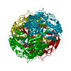
 Structure visualization
 Downloads & links
 Other downloads
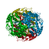

 PDBj
PDBj


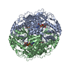
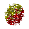
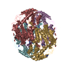
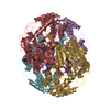
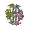
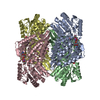
 Assembly


 Mass: 173.040 Da / Num. of mol.: 1 / Source method: obtained synthetically / Formula: Yb
 Mass: 35.453 Da / Num. of mol.: 1 / Source method: obtained synthetically / Formula: Cl
 Mass: 18.015 Da / Num. of mol.: 246 / Source method: isolated from a natural source / Formula: H2O
 Sample preparation
 Processing