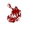
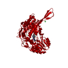
 Movie
Movie Controller


+ Open data

- Basic information
| Entry | Database: PDB / ID: 1o05 | ||||||
|---|---|---|---|---|---|---|---|
| Title | Apo form of human mitochondrial aldehyde dehydrogenase | ||||||
 Components Components | Aldehyde dehydrogenase | ||||||
 Keywords Keywords | OXIDOREDUCTASE / ALDH / NAD / NADH / isomerization | ||||||
| Function / homology |  Function and homology information Function and homology informationMetabolism of serotonin / nitroglycerin metabolic process / regulation of dopamine biosynthetic process / regulation of serotonin biosynthetic process / phenylacetaldehyde dehydrogenase (NAD+) activity / ethanol metabolic process / aldehyde catabolic process / alcohol metabolic process / ethanol catabolic process / Ethanol oxidation ...Metabolism of serotonin / nitroglycerin metabolic process / regulation of dopamine biosynthetic process / regulation of serotonin biosynthetic process / phenylacetaldehyde dehydrogenase (NAD+) activity / ethanol metabolic process / aldehyde catabolic process / alcohol metabolic process / ethanol catabolic process / Ethanol oxidation / aldehyde dehydrogenase [NAD(P)+] activity / aldehyde dehydrogenase (NAD+) / carboxylesterase activity / cellular detoxification of aldehyde / aldehyde dehydrogenase (NAD+) activity / Smooth Muscle Contraction / Mitochondrial protein degradation / NAD binding / carbohydrate metabolic process / electron transfer activity / mitochondrial matrix / mitochondrion / extracellular exosome Similarity search - Function | ||||||
| Biological species |  Homo sapiens (human) Homo sapiens (human) | ||||||
| Method |  X-RAY DIFFRACTION / MOLECULAR REPLACEMENT / Resolution: 2.25 Å X-RAY DIFFRACTION / MOLECULAR REPLACEMENT / Resolution: 2.25 Å | ||||||
 Authors Authors | Hurley, T.D. / Perez-Miller, S. / Breen, H. | ||||||
 Citation Citation | Journal: Chem.Biol.Interact. / Year: 2001 Title: Order and disorder in mitochondrial aldehyde dehydrogenase Authors: Hurley, T.D. / Perez-Miller, S. / Breen, H. #1: Journal: Biochemistry / Year: 2003Title: Coenzyme isomerization is integral to catalysis in aldehyde dehydrogenase Authors: Perez-Miller, S.J. / Hurley, T.D. | ||||||
| History |
|
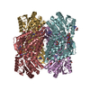
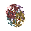
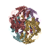
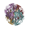
- Structure visualization
| Structure viewer | Molecule: MolmilJmol/JSmol |
|---|
- Downloads & links
-Download
| PDBx/mmCIF format | 1o05.cif.gz | 803.6 KB | Display | PDBx/mmCIF format |
|---|---|---|---|---|
| PDB format | pdb1o05.ent.gz | 660.4 KB | Display | PDB format |
| PDBx/mmJSON format | 1o05.json.gz | Tree view | PDBx/mmJSON format | |
| Others |  Other downloads Other downloads |
-Validation report
| Arichive directory | https://data.pdbj.org/pub/pdb/validation_reports/o0/1o05ftp://data.pdbj.org/pub/pdb/validation_reports/o0/1o05 | HTTPS FTP |
|---|
-Related structure data
| Related structure data | 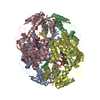 1cw3S S: Starting model for refinement |
|---|---|
| Similar structure data |


-Links
 PDBj
PDBj


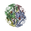
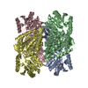
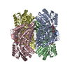
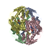
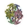
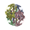
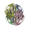
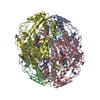
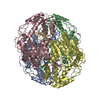
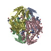
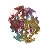
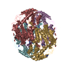
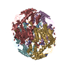
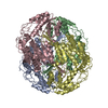
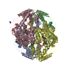
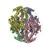
- Assembly
| Deposited unit | 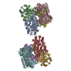
| ||||||||
|---|---|---|---|---|---|---|---|---|---|
| 1 | 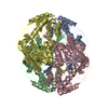
| ||||||||
| 2 | 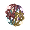
| ||||||||
| Unit cell |
|
-Components
| #1: Protein | Mass: 54499.629 Da / Num. of mol.: 8 Fragment: Complete mature sequence (does not contain mitochondrial leader sequence). Source method: isolated from a genetically manipulated source Source: (gene. exp.) Homo sapiens (human) / Gene: ALDH2 OR ALDM / Plasmid: PET7-7 / Species (production host): Escherichia coli / Production host:  #2: Chemical | ChemComp-NA /   Mass: 22.990 Da / Num. of mol.: 8 / Source method: obtained synthetically / Formula: Na Mass: 22.990 Da / Num. of mol.: 8 / Source method: obtained synthetically / Formula: Na#3: Water | ChemComp-HOH / |  Mass: 18.015 Da / Num. of mol.: 3210 / Source method: isolated from a natural source / Formula: H2O Mass: 18.015 Da / Num. of mol.: 3210 / Source method: isolated from a natural source / Formula: H2O |
|---|
-Experimental details
-Experiment
| Experiment | Method: X-RAY DIFFRACTION / Number of used crystals: 1 |
|---|
- Sample preparation
| Crystal | Density Matthews: 2.02 Å3/Da / Density % sol: 38.6 % | |||||||||||||||||||||||||||||||||||
|---|---|---|---|---|---|---|---|---|---|---|---|---|---|---|---|---|---|---|---|---|---|---|---|---|---|---|---|---|---|---|---|---|---|---|---|---|
| Crystal grow | Temperature: 293 K / Method: vapor diffusion, sitting drop / pH: 6.4 Details: ACES, PEG 6000, Guanidine HCl, MgCl2, DTT., pH 6.4, VAPOR DIFFUSION, SITTING DROP, temperature 293K | |||||||||||||||||||||||||||||||||||
| Crystal grow | *PLUS pH: 6.2 / Method: unknown / Details: Ni, L., (1999) Protein Sci., 8, 2784. | |||||||||||||||||||||||||||||||||||
| Components of the solutions | *PLUS
|
-Data collection
| Diffraction | Mean temperature: 110 K |
|---|---|
| Diffraction source | Source: ROTATING ANODE / Type: RIGAKU RU200 / Wavelength: 1.5418 Å |
| Detector | Type: RIGAKU RAXIS IIC / Detector: IMAGE PLATE / Date: Nov 3, 1999 |
| Radiation | Monochromator: Yale mirrors / Protocol: SINGLE WAVELENGTH / Monochromatic (M) / Laue (L): M / Scattering type: x-ray |
| Radiation wavelength | Wavelength: 1.5418 Å / Relative weight: 1 |
| Reflection | Resolution: 2.25→25 Å / Num. all: 181352 / Num. obs: 165756 / % possible obs: 91.4 % / Observed criterion σ(F): 0 / Observed criterion σ(I): 0 / Redundancy: 3 % / Biso Wilson estimate: 12.6 Å2 / Rmerge(I) obs: 0.072 / Net I/σ(I): 14.3 |
| Reflection shell | Resolution: 2.25→2.33 Å / Rmerge(I) obs: 0.214 / Mean I/σ(I) obs: 3.5 / Num. unique all: 11115 / % possible all: 62 |
| Reflection | *PLUS Lowest resolution: 42 Å / Num. measured all: 500412 |
| Reflection shell | *PLUS % possible obs: 62 % |
- Processing
| Software |
| ||||||||||||||||||||||||||||||||||||
|---|---|---|---|---|---|---|---|---|---|---|---|---|---|---|---|---|---|---|---|---|---|---|---|---|---|---|---|---|---|---|---|---|---|---|---|---|---|
| Refinement | Method to determine structure: MOLECULAR REPLACEMENT Starting model: 1CW3 Resolution: 2.25→24.96 Å / Rfactor Rfree error: 0.002 / Data cutoff high absF: 5593690.14 / Data cutoff high rms absF: 5593690.14 / Data cutoff low absF: 0 / Isotropic thermal model: RESTRAINED / Cross valid method: THROUGHOUT / σ(F): 0 / σ(I): 0 / Stereochemistry target values: Engh & Huber / Details: maximum likelihood target using amplitudes
| ||||||||||||||||||||||||||||||||||||
| Solvent computation | Solvent model: FLAT MODEL / Bsol: 10 Å2 / ksol: 0.299682 e/Å3 | ||||||||||||||||||||||||||||||||||||
| Displacement parameters | Biso mean: 16.8 Å2
| ||||||||||||||||||||||||||||||||||||
| Refine analyze |
| ||||||||||||||||||||||||||||||||||||
| Refinement step | Cycle: LAST / Resolution: 2.25→24.96 Å
| ||||||||||||||||||||||||||||||||||||
| Refine LS restraints |
| ||||||||||||||||||||||||||||||||||||
| LS refinement shell | Resolution: 2.25→2.33 Å / Rfactor Rfree error: 0.011 / Total num. of bins used: 10
| ||||||||||||||||||||||||||||||||||||
| Xplor file |
| ||||||||||||||||||||||||||||||||||||
| Refinement | *PLUS Lowest resolution: 42 Å / Rfactor Rfree: 0.226 / Rfactor Rwork: 0.176 | ||||||||||||||||||||||||||||||||||||
| Solvent computation | *PLUS | ||||||||||||||||||||||||||||||||||||
| Displacement parameters | *PLUS | ||||||||||||||||||||||||||||||||||||
| Refine LS restraints | *PLUS
|
