 Movie
Movie Controller

[English] 日本語
 Yorodumi
- PDB-2ono: Arg475Gln Mutant of Mitochondrial Aldehyde Dehydrogenase, apo for... -
+ Open data

- Basic information
| Entry | Database: PDB / ID: 2ono | ||||||
|---|---|---|---|---|---|---|---|
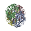
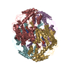
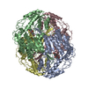
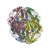
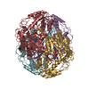
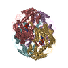
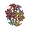
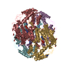
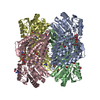
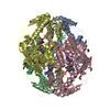
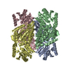
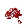
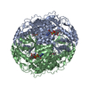
| Title | Arg475Gln Mutant of Mitochondrial Aldehyde Dehydrogenase, apo form, pseudo-merohedrally twinned | ||||||
 Components Components | Aldehyde dehydrogenase | ||||||
 Keywords Keywords | OXIDOREDUCTASE / ALDH / NAD / Rossmann Fold | ||||||
| Function / homology |  Function and homology information Function and homology informationMetabolism of serotonin / nitroglycerin metabolic process / regulation of dopamine biosynthetic process / regulation of serotonin biosynthetic process / phenylacetaldehyde dehydrogenase (NAD+) activity / ethanol metabolic process / aldehyde catabolic process / alcohol metabolic process / ethanol catabolic process / Ethanol oxidation ...Metabolism of serotonin / nitroglycerin metabolic process / regulation of dopamine biosynthetic process / regulation of serotonin biosynthetic process / phenylacetaldehyde dehydrogenase (NAD+) activity / ethanol metabolic process / aldehyde catabolic process / alcohol metabolic process / ethanol catabolic process / Ethanol oxidation / aldehyde dehydrogenase [NAD(P)+] activity / aldehyde dehydrogenase (NAD+) / carboxylesterase activity / cellular detoxification of aldehyde / aldehyde dehydrogenase (NAD+) activity / Smooth Muscle Contraction / Mitochondrial protein degradation / NAD binding / carbohydrate metabolic process / electron transfer activity / mitochondrial matrix / mitochondrion / extracellular exosome Similarity search - Function | ||||||
| Biological species |  Homo sapiens (human) Homo sapiens (human) | ||||||
| Method |  X-RAY DIFFRACTION / SYNCHROTRON / MOLECULAR REPLACEMENT / Resolution: 2.15 Å X-RAY DIFFRACTION / SYNCHROTRON / MOLECULAR REPLACEMENT / Resolution: 2.15 Å | ||||||
 Authors Authors | Larson, H.N. / Hurley, T.D. | ||||||
 Citation Citation | Journal: J.Biol.Chem. / Year: 2007 Title: Structural and functional consequences of coenzyme binding to the inactive asian variant of mitochondrial aldehyde dehydrogenase: roles of residues 475 and 487. Authors: Larson, H.N. / Zhou, J. / Chen, Z. / Stamler, J.S. / Weiner, H. / Hurley, T.D. | ||||||
| History |
|
- Structure visualization
| Structure viewer | Molecule: MolmilJmol/JSmol |
|---|
- Downloads & links
-Download
| PDBx/mmCIF format | 2ono.cif.gz | 744.7 KB | Display | PDBx/mmCIF format |
|---|---|---|---|---|
| PDB format | pdb2ono.ent.gz | 617.1 KB | Display | PDB format |
| PDBx/mmJSON format | 2ono.json.gz | Tree view | PDBx/mmJSON format | |
| Others |  Other downloads Other downloads |
-Validation report
| Arichive directory | https://data.pdbj.org/pub/pdb/validation_reports/on/2onoftp://data.pdbj.org/pub/pdb/validation_reports/on/2ono | HTTPS FTP |
|---|
-Related structure data
| Related structure data | 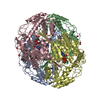 2onmC 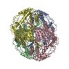 2onnC  2onpC 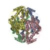 1o05S S: Starting model for refinement C: citing same article ( |
|---|---|
| Similar structure data |


-Links
 PDBj
PDBj


- Assembly
| Deposited unit | 
| ||||||||
|---|---|---|---|---|---|---|---|---|---|
| 1 | 
| ||||||||
| 2 | 
| ||||||||
| Unit cell |
| ||||||||
| Details | The biological unit is a tetramer. There are 3 biological untils in the assymmetric unit. The biological unit 1 consists of chain(s) A, B, C, D. The biological unit 2 consists of chain(s) E, F, G, H. |
-Components
| #1: Protein | Mass: 54470.562 Da / Num. of mol.: 8 / Mutation: R475Q Source method: isolated from a genetically manipulated source Source: (gene. exp.) Homo sapiens (human) / Gene: ALDH2, ALDM / Plasmid: pT-7-7 / Species (production host): Escherichia coli / Production host:  #2: Water | ChemComp-HOH / |  Mass: 18.015 Da / Num. of mol.: 1020 / Source method: isolated from a natural source / Formula: H2O Mass: 18.015 Da / Num. of mol.: 1020 / Source method: isolated from a natural source / Formula: H2O |
|---|
-Experimental details
-Experiment
| Experiment | Method: X-RAY DIFFRACTION / Number of used crystals: 1 |
|---|
- Sample preparation
| Crystal | Density Matthews: 2.069 Å3/Da / Density % sol: 41.15 % |
|---|---|
| Crystal grow | Temperature: 293 K / pH: 6.6 Details: 100 mM ACES (N-[2-Acetamido]-2-aminoethane sufonic acid), 5 mM MgCl2, 100 mM Guanidine HCl, 18% w/v PEG 6000, 6 mM DTT, pH 6.6, vapor diffusion, temperature 293K, pH 6.60 |
-Data collection
| Diffraction | Mean temperature: 100 K |
|---|---|
| Diffraction source | Source: SYNCHROTRON / Site: APS  / Beamline: 14-BM-C / Wavelength: 0.9 / Beamline: 14-BM-C / Wavelength: 0.9 |
| Detector | Type: ADSC QUANTUM 4 / Detector: CCD / Date: Aug 3, 2003 |
| Radiation | Monochromator: SI (111) / Protocol: SINGLE WAVELENGTH / Monochromatic (M) / Laue (L): M / Scattering type: x-ray |
| Radiation wavelength | Wavelength: 0.9 Å / Relative weight: 1 |
| Reflection | Resolution: 2.15→25 Å / Num. obs: 186854 / % possible obs: 97.5 % / Observed criterion σ(I): 0.2 / Biso Wilson estimate: 21.84 Å2 / Rmerge(I) obs: 0.061 / Net I/σ(I): 13.6 |
| Reflection shell | Resolution: 2.15→2.23 Å / Rmerge(I) obs: 0.198 / % possible all: 87.8 |
- Processing
| Software |
| |||||||||||||||||||||||||||||||||
|---|---|---|---|---|---|---|---|---|---|---|---|---|---|---|---|---|---|---|---|---|---|---|---|---|---|---|---|---|---|---|---|---|---|---|
| Refinement | Method to determine structure: MOLECULAR REPLACEMENT Starting model: PDB ENTRY 1O05 Resolution: 2.15→10 Å / Num. parameters: 125536 / Num. restraintsaints: 316424 / Cross valid method: FREE R / σ(F): 0 / Stereochemistry target values: ENGH & HUBER
| |||||||||||||||||||||||||||||||||
| Refine analyze | Num. disordered residues: 0 / Occupancy sum hydrogen: 0 / Occupancy sum non hydrogen: 31383 | |||||||||||||||||||||||||||||||||
| Refinement step | Cycle: LAST / Resolution: 2.15→10 Å
| |||||||||||||||||||||||||||||||||
| Refine LS restraints |
|
