 Movie
Movie Controller

[English] 日本語
 Yorodumi
- PDB-3sz9: Crystal structure of human ALDH2 modified with the beta-eliminati... -
+ Open data

- Basic information
| Entry | Database: PDB / ID: 3sz9 | ||||||
|---|---|---|---|---|---|---|---|
| Title | Crystal structure of human ALDH2 modified with the beta-elimination product of Aldi-3; 1-(4-ethylbenzene)prop-2-en-1-one | ||||||
 Components Components | Aldehyde dehydrogenase, mitochondrial | ||||||
 Keywords Keywords | OXIDOREDUCTASE/OXIDOREDUCTASE INHIBITOR / ALDH / Aldi-3 / inhibitor / Rossmann Fold / Oxidoreductase / Covalent adduct / Mitochondria / OXIDOREDUCTASE-OXIDOREDUCTASE INHIBITOR complex | ||||||
| Function / homology |  Function and homology information Function and homology informationMetabolism of serotonin / nitroglycerin metabolic process / regulation of dopamine biosynthetic process / regulation of serotonin biosynthetic process / phenylacetaldehyde dehydrogenase (NAD+) activity / ethanol metabolic process / aldehyde catabolic process / alcohol metabolic process / ethanol catabolic process / Ethanol oxidation ...Metabolism of serotonin / nitroglycerin metabolic process / regulation of dopamine biosynthetic process / regulation of serotonin biosynthetic process / phenylacetaldehyde dehydrogenase (NAD+) activity / ethanol metabolic process / aldehyde catabolic process / alcohol metabolic process / ethanol catabolic process / Ethanol oxidation / aldehyde dehydrogenase [NAD(P)+] activity / aldehyde dehydrogenase (NAD+) / carboxylesterase activity / cellular detoxification of aldehyde / aldehyde dehydrogenase (NAD+) activity / Smooth Muscle Contraction / Mitochondrial protein degradation / NAD binding / carbohydrate metabolic process / electron transfer activity / mitochondrial matrix / mitochondrion / extracellular exosome Similarity search - Function | ||||||
| Biological species |  Homo sapiens (human) Homo sapiens (human) | ||||||
| Method |  X-RAY DIFFRACTION / SYNCHROTRON / MOLECULAR REPLACEMENT / Resolution: 2.1 Å X-RAY DIFFRACTION / SYNCHROTRON / MOLECULAR REPLACEMENT / Resolution: 2.1 Å | ||||||
 Authors Authors | Perez-Miller, S. / Hurley, T.D. | ||||||
 Citation Citation | Journal: J.Biol.Chem. / Year: 2011 Title: Discovery of a novel class of covalent inhibitor for aldehyde dehydrogenases. Authors: Khanna, M. / Chen, C.H. / Kimble-Hill, A. / Parajuli, B. / Perez-Miller, S. / Baskaran, S. / Kim, J. / Dria, K. / Vasiliou, V. / Mochly-Rosen, D. / Hurley, T.D. #1: Journal: Nat.Struct.Mol.Biol. / Year: 2010Title: Alda-1 is an agonist and chemical chaperone for the common human aldehyde dehydrogenase 2 variant Authors: Perez-Miller, S. / Younus, H. / Vanam, R. / Chen, C.-H. / Mochly-Rosen, D. / Hurley, T.D. #2: Journal: Science / Year: 2008Title: Activation of Aldehyde Dehydrogenase-2 Reduces Ischemic Damage to the Heart Authors: Chen, C.-H. / Budas, G.R. / Churchill, E.N. / Disatnik, M.-H. / Hurley, T.D. / Mochly-Rosen, D. | ||||||
| History |
|
- Structure visualization
| Structure viewer | Molecule: MolmilJmol/JSmol |
|---|
- Downloads & links
-Download
| PDBx/mmCIF format | 3sz9.cif.gz | 803.6 KB | Display | PDBx/mmCIF format |
|---|---|---|---|---|
| PDB format | pdb3sz9.ent.gz | 662.9 KB | Display | PDB format |
| PDBx/mmJSON format | 3sz9.json.gz | Tree view | PDBx/mmJSON format | |
| Others |  Other downloads Other downloads |
-Validation report
| Arichive directory | https://data.pdbj.org/pub/pdb/validation_reports/sz/3sz9ftp://data.pdbj.org/pub/pdb/validation_reports/sz/3sz9 | HTTPS FTP |
|---|
-Related structure data
| Related structure data |  3szaC  3szbC  1o05S C: citing same article ( S: Starting model for refinement |
|---|---|
| Similar structure data |


-Links
 PDBj
PDBj


- Assembly
| Deposited unit | 
| ||||||||
|---|---|---|---|---|---|---|---|---|---|
| 1 | 
| ||||||||
| 2 | 
| ||||||||
| 3 | 
| ||||||||
| 4 | 
| ||||||||
| 5 | 
| ||||||||
| 6 | 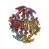
| ||||||||
| Unit cell |
| ||||||||
| Details | There are two tetramers in the asymmetric unit (chains A, B, C, & D and chains E, F, G & H) |
-Components
-Protein , 1 types, 8 molecules ABCDEFGH
| #1: Protein | Mass: 54499.629 Da / Num. of mol.: 8 / Fragment: Mature sequence, UNP residues 18-517 Source method: isolated from a genetically manipulated source Details: lacks mitochondrial leader sequence / Source: (gene. exp.) Homo sapiens (human) / Gene: ALDH2, ALDM / Plasmid: pT-7-7 / Production host:  |
|---|
-Non-polymers , 5 types, 2649 molecules 


| #2: Chemical | ChemComp-NA /  Mass: 22.990 Da / Num. of mol.: 8 / Source method: obtained synthetically / Formula: Na Mass: 22.990 Da / Num. of mol.: 8 / Source method: obtained synthetically / Formula: Na#3: Chemical | ChemComp-GAI /  Mass: 59.070 Da / Num. of mol.: 18 / Source method: obtained synthetically / Formula: CH5N3 Mass: 59.070 Da / Num. of mol.: 18 / Source method: obtained synthetically / Formula: CH5N3#4: Chemical | ChemComp-EDO /  Mass: 62.068 Da / Num. of mol.: 21 / Source method: obtained synthetically / Formula: C2H6O2 Mass: 62.068 Da / Num. of mol.: 21 / Source method: obtained synthetically / Formula: C2H6O2#5: Chemical | ChemComp-I3E /  Mass: 162.228 Da / Num. of mol.: 8 / Source method: obtained synthetically / Formula: C11H14O Mass: 162.228 Da / Num. of mol.: 8 / Source method: obtained synthetically / Formula: C11H14O#6: Water | ChemComp-HOH / | Mass: 18.015 Da / Num. of mol.: 2594 / Source method: isolated from a natural source / Formula: H2O |
|---|
-Details
| Has protein modification | Y |
|---|---|
| Nonpolymer details | THE STARTING MATERIAL IS 1-(4-ETHYLPHENYL)PROP-2-EN-1-ONE. IT BINDS COVALENTLY TO CYS 302. I3E ...THE STARTING MATERIAL IS 1-(4-ETHYLPHENY |
-Experimental details
-Experiment
| Experiment | Method: X-RAY DIFFRACTION / Number of used crystals: 1 |
|---|
- Sample preparation
| Crystal | Density Matthews: 2.15 Å3/Da / Density % sol: 42.91 % |
|---|---|
| Crystal grow | Temperature: 292 K / Method: vapor diffusion / pH: 6.4 Details: 100 MM ACES (N-[2-ACETAMIDO]-2-AMINOETHANE SULFONIC ACID), 10MM MGCL2, 100 MM GUANIDINE HCL, 16-17% W/V PEG 6000, 4MM DTT, pH 6.4, vapor diffusion, temperature 292K |
-Data collection
| Diffraction | Mean temperature: 100 K | |||||||||||||||||||||||||||||||||||||||||||||||||||||||||||||||||||||||||||||||||||||||||||||||||||||||||||||||||||||||||||||||||||||||||||||||||||
|---|---|---|---|---|---|---|---|---|---|---|---|---|---|---|---|---|---|---|---|---|---|---|---|---|---|---|---|---|---|---|---|---|---|---|---|---|---|---|---|---|---|---|---|---|---|---|---|---|---|---|---|---|---|---|---|---|---|---|---|---|---|---|---|---|---|---|---|---|---|---|---|---|---|---|---|---|---|---|---|---|---|---|---|---|---|---|---|---|---|---|---|---|---|---|---|---|---|---|---|---|---|---|---|---|---|---|---|---|---|---|---|---|---|---|---|---|---|---|---|---|---|---|---|---|---|---|---|---|---|---|---|---|---|---|---|---|---|---|---|---|---|---|---|---|---|---|---|---|
| Diffraction source | Source: SYNCHROTRON / Site: APS  / Beamline: 19-ID / Wavelength: 0.9869 Å / Beamline: 19-ID / Wavelength: 0.9869 Å | |||||||||||||||||||||||||||||||||||||||||||||||||||||||||||||||||||||||||||||||||||||||||||||||||||||||||||||||||||||||||||||||||||||||||||||||||||
| Detector | Type: ADSC QUANTUM 315 / Detector: CCD / Date: Apr 28, 2009 | |||||||||||||||||||||||||||||||||||||||||||||||||||||||||||||||||||||||||||||||||||||||||||||||||||||||||||||||||||||||||||||||||||||||||||||||||||
| Radiation | Monochromator: SAGITALLY FOCUSED Si(111) / Protocol: SINGLE WAVELENGTH / Monochromatic (M) / Laue (L): M / Scattering type: x-ray | |||||||||||||||||||||||||||||||||||||||||||||||||||||||||||||||||||||||||||||||||||||||||||||||||||||||||||||||||||||||||||||||||||||||||||||||||||
| Radiation wavelength | Wavelength: 0.9869 Å / Relative weight: 1 | |||||||||||||||||||||||||||||||||||||||||||||||||||||||||||||||||||||||||||||||||||||||||||||||||||||||||||||||||||||||||||||||||||||||||||||||||||
| Reflection | Resolution: 2.1→50 Å / Num. obs: 217889 / % possible obs: 99.7 % / Observed criterion σ(I): 0.2 / Redundancy: 5.5 % / Biso Wilson estimate: 31.6 Å2 / Rmerge(I) obs: 0.098 / Χ2: 1.064 / Net I/σ(I): 7.5 | |||||||||||||||||||||||||||||||||||||||||||||||||||||||||||||||||||||||||||||||||||||||||||||||||||||||||||||||||||||||||||||||||||||||||||||||||||
| Reflection shell |
|
- Processing
| Software |
| |||||||||||||||||||||||||||||||||||||||||||||||||||||||||||||||||
|---|---|---|---|---|---|---|---|---|---|---|---|---|---|---|---|---|---|---|---|---|---|---|---|---|---|---|---|---|---|---|---|---|---|---|---|---|---|---|---|---|---|---|---|---|---|---|---|---|---|---|---|---|---|---|---|---|---|---|---|---|---|---|---|---|---|---|
| Refinement | Method to determine structure: MOLECULAR REPLACEMENT Starting model: PDB Entry 1O05 Resolution: 2.1→50 Å / Cor.coef. Fo:Fc: 0.96 / Cor.coef. Fo:Fc free: 0.938 / Occupancy max: 1 / Occupancy min: 0.3 / SU B: 5.077 / SU ML: 0.136 / SU R Cruickshank DPI: 0 / Isotropic thermal model: Isotropic / Cross valid method: THROUGHOUT / ESU R Free: 0.19 / Stereochemistry target values: MAXIMUM LIKELIHOOD Details: HYDROGENS HAVE BEEN ADDED IN THE RIDING POSITIONS U VALUES : REFINED INDIVIDUALLY
| |||||||||||||||||||||||||||||||||||||||||||||||||||||||||||||||||
| Solvent computation | Ion probe radii: 0.8 Å / Shrinkage radii: 0.8 Å / VDW probe radii: 1.4 Å / Solvent model: BABINET MODEL WITH MASK | |||||||||||||||||||||||||||||||||||||||||||||||||||||||||||||||||
| Displacement parameters | Biso max: 78.92 Å2 / Biso mean: 26.0324 Å2 / Biso min: 6.69 Å2
| |||||||||||||||||||||||||||||||||||||||||||||||||||||||||||||||||
| Refinement step | Cycle: LAST / Resolution: 2.1→50 Å
| |||||||||||||||||||||||||||||||||||||||||||||||||||||||||||||||||
| Refine LS restraints |
| |||||||||||||||||||||||||||||||||||||||||||||||||||||||||||||||||
| LS refinement shell | Resolution: 2.101→2.155 Å / Total num. of bins used: 20
|
