 Movie
Movie Controller

[English] 日本語
 Yorodumi
- PDB-1lvn: CRYSTAL STRUCTURE OF E. COLI AMINE OXIDASE COMPLEXED WITH TRANYLC... -
+ Open data

- Basic information
| Entry | Database: PDB / ID: 1lvn | |||||||||
|---|---|---|---|---|---|---|---|---|---|---|
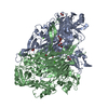
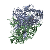
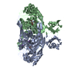
| Title | CRYSTAL STRUCTURE OF E. COLI AMINE OXIDASE COMPLEXED WITH TRANYLCYPROMINE | |||||||||
 Components Components | Copper amine oxidase | |||||||||
 Keywords Keywords | OXIDOREDUCTASE / Inhibitor complex | |||||||||
| Function / homology |  Function and homology information Function and homology informationphenylethylamine catabolic process / primary-amine oxidase / primary methylamine oxidase activity / amine metabolic process / L-phenylalanine catabolic process / quinone binding / periplasmic space / copper ion binding / calcium ion binding Similarity search - Function | |||||||||
| Biological species |  | |||||||||
| Method |  X-RAY DIFFRACTION / SYNCHROTRON / FOURIER SYNTHESIS / Resolution: 2.4 Å X-RAY DIFFRACTION / SYNCHROTRON / FOURIER SYNTHESIS / Resolution: 2.4 Å | |||||||||
 Authors Authors | Wilmot, C.M. / Phillips, S.E. | |||||||||
 Citation Citation | Journal: Febs Lett. / Year: 2004 Title: Medical implications from the crystal structure of a copper-containing amine oxidase complexed with the antidepressant drug tranylcypromine. Authors: Wilmot, C.M. / Saysell, C.G. / Blessington, A. / Conn, D.A. / Kurtis, C.R. / McPherson, M.J. / Knowles, P.F. / Phillips, S.E. #1: Journal: BIOCHEM.J. / Year: 2002Title: Probing the catalytic mechanism of Escherichia coli amine oxidase using mutational variants and a reversible inhibitor as a substrate analogue. Authors: Saysell, C.G. / Tambyrajah, W.S. / Murray, J.M. / Wilmot, C.M. / Phillips, S.E. / McPherson, M.J. / Knowles, P.F. #2: Journal: Biochemistry / Year: 1997Title: Catalytic Mechanism of the Quinoenzyme Amine Oxidase from Escherichia Coli: Exploring the Reductive Half-Reaction. Authors: Wilmot, C.M. / Murray, J.M. / Alton, G. / Parsons, M.R. / Convery, M.A. / Blakeley, V. / Corner, A.S. / Palcic, M.M. / Knowles, P.F. / McPherson, M.J. / Phillips, S.E.V. #3: Journal: Structure / Year: 1995Title: Crystal Structure of a Quinoenzyme: Copper Amine Oxidase of Escherichia Coli at 2 Angstroms Resolution. Authors: Parsons, M.R. / Convery, M.A. / Wilmot, C.M. / Yadav, K.D.S. / Blakeley, V. / Corner, A.S. / Phillips, S.E. / McPherson, M.J. / Knowles, P.F. #4: Journal: Biochemistry / Year: 1999Title: The active site base controls cofactor reactivity in Escherichia coli amine oxidase: X-ray crystallographic studies with mutational variants. Authors: Murray, J.M. / Saysell, C.G. / Wilmot, C.M. / Tambyrajah, W.S. / Jaeger, J. / Knowles, P.F. / Phillips, S.E. / McPherson, M.J. #5: Journal: Science / Year: 1999Title: Visualization of dioxygen bound to copper during enzyme catalysis. Authors: Wilmot, C.M. / Hajdu, J. / McPherson, M.J. / Knowles, P.F. / Phillips, S.E. | |||||||||
| History |
|
- Structure visualization
| Structure viewer | Molecule: MolmilJmol/JSmol |
|---|
- Downloads & links
-Download
| PDBx/mmCIF format | 1lvn.cif.gz | 326 KB | Display | PDBx/mmCIF format |
|---|---|---|---|---|
| PDB format | pdb1lvn.ent.gz | 262 KB | Display | PDB format |
| PDBx/mmJSON format | 1lvn.json.gz | Tree view | PDBx/mmJSON format | |
| Others |  Other downloads Other downloads |
-Validation report
| Arichive directory | https://data.pdbj.org/pub/pdb/validation_reports/lv/1lvnftp://data.pdbj.org/pub/pdb/validation_reports/lv/1lvn | HTTPS FTP |
|---|
-Related structure data
| Related structure data |  1spuS S: Starting model for refinement |
|---|---|
| Similar structure data |


-Links
 PDBj
PDBj- Assembly
| Deposited unit | 
| ||||||||
|---|---|---|---|---|---|---|---|---|---|
| 1 |
| ||||||||
| Unit cell |
|
-Components
| #1: Protein | Mass: 81483.922 Da / Num. of mol.: 2 Source method: isolated from a genetically manipulated source Source: (gene. exp.) #2: Chemical |   Mass: 63.546 Da / Num. of mol.: 2 / Source method: obtained synthetically / Formula: Cu Mass: 63.546 Da / Num. of mol.: 2 / Source method: obtained synthetically / Formula: Cu#3: Chemical | ChemComp-CA /   Mass: 40.078 Da / Num. of mol.: 4 / Source method: obtained synthetically / Formula: Ca Mass: 40.078 Da / Num. of mol.: 4 / Source method: obtained synthetically / Formula: Ca#4: Water | ChemComp-HOH / |  Mass: 18.015 Da / Num. of mol.: 1447 / Source method: isolated from a natural source / Formula: H2O Mass: 18.015 Da / Num. of mol.: 1447 / Source method: isolated from a natural source / Formula: H2OHas protein modification | Y | |
|---|
-Experimental details
-Experiment
| Experiment | Method: X-RAY DIFFRACTION / Number of used crystals: 1 |
|---|
- Sample preparation
| Crystal | Density Matthews: 2.76 Å3/Da / Density % sol: 0.55 % | |||||||||||||||
|---|---|---|---|---|---|---|---|---|---|---|---|---|---|---|---|---|
| Crystal grow | Temperature: 291 K / Method: sitting drop / pH: 7.2 Details: 1.3M SODIUM CITRATE, 0.1M HEPES BUFFER, pH 7.2, INHIBITOR SOAKING SOLUTION 10:1 RATIO OF RACEMIC TRANYLCYPROMINE TO ENZYME MADE UP IN 1.4M SODIUM CITRATE, 0.1M HEPES BUFFER, PH 7.2. CRYSTAL ...Details: 1.3M SODIUM CITRATE, 0.1M HEPES BUFFER, pH 7.2, INHIBITOR SOAKING SOLUTION 10:1 RATIO OF RACEMIC TRANYLCYPROMINE TO ENZYME MADE UP IN 1.4M SODIUM CITRATE, 0.1M HEPES BUFFER, PH 7.2. CRYSTAL SOAKED FOR 20 DAYS.CRYOPROTECTANT 20% GLYCEROL, 1.4M SODIUM CITRATE BUFFER, PH 7.2, pH 7.20, Sitting drop, temperature 291.0K | |||||||||||||||
| Crystal grow | *PLUS pH: 7 / Method: vapor diffusion / Details: Parsons, M.R., (1995) Structure, 3, 1171. | |||||||||||||||
| Components of the solutions | *PLUS
|
-Data collection
| Diffraction | Mean temperature: 100 K |
|---|---|
| Diffraction source | Source: SYNCHROTRON / Site: SRS  / Beamline: PX9.6 / Wavelength: 0.87 / Wavelength: 0.87 Å / Beamline: PX9.6 / Wavelength: 0.87 / Wavelength: 0.87 Å |
| Detector | Type: MAR scanner 300 mm plate / Detector: IMAGE PLATE / Date: Oct 20, 1996 |
| Radiation | Protocol: SINGLE WAVELENGTH / Monochromatic (M) / Laue (L): M / Scattering type: x-ray |
| Radiation wavelength | Wavelength: 0.87 Å / Relative weight: 1 |
| Reflection | Resolution: 2.4→100 Å / Num. all: 64108 / Num. obs: 64108 / % possible obs: 90.1 % / Observed criterion σ(I): 0 / Redundancy: 2.56 % / Biso Wilson estimate: 36.6 Å2 / Rmerge(I) obs: 0.075 / Net I/σ(I): 7.5 |
| Reflection shell | Resolution: 2.4→2.49 Å / Redundancy: 2.1 % / Rmerge(I) obs: 0.251 / Mean I/σ(I) obs: 2.9 / % possible all: 80.6 |
| Reflection | *PLUS Highest resolution: 2.4 Å / Lowest resolution: 20 Å / Num. obs: 64108 / % possible obs: 90.1 % / Num. measured all: 163856 / Rmerge(I) obs: 0.075 |
| Reflection shell | *PLUS % possible obs: 80.6 % / Rmerge(I) obs: 0.251 / Mean I/σ(I) obs: 2.9 |
- Processing
| Software |
| ||||||||||||||||||||||||||||
|---|---|---|---|---|---|---|---|---|---|---|---|---|---|---|---|---|---|---|---|---|---|---|---|---|---|---|---|---|---|
| Refinement | Method to determine structure: FOURIER SYNTHESIS Starting model: 1SPU Resolution: 2.4→20 Å / Cross valid method: FREE R / σ(F): 0 / Stereochemistry target values: ENGH AND HUBER Details: CYCLES OF POSITIONAL POWELL MINIMIZATION AND INDIVIDUAL TEMPERATURE FACTOR REFINEMENT, WITH A MLF TARGET, AND AN OVERALL ANISOTROPIC TEMPERATURE FACTOR CORRECTION. COPPER ION RESTRAINTS WERE ...Details: CYCLES OF POSITIONAL POWELL MINIMIZATION AND INDIVIDUAL TEMPERATURE FACTOR REFINEMENT, WITH A MLF TARGET, AND AN OVERALL ANISOTROPIC TEMPERATURE FACTOR CORRECTION. COPPER ION RESTRAINTS WERE WEAKENED BY ADJUSTMENT OF THE NON-BONDED PARAMTERES, TO ENABLE THE COPPER ION/LIGAND DISTANCES TO REFLECT THE DATA MORE ACCURATELY.
| ||||||||||||||||||||||||||||
| Displacement parameters |
| ||||||||||||||||||||||||||||
| Refinement step | Cycle: LAST / Resolution: 2.4→20 Å
| ||||||||||||||||||||||||||||
| Refine LS restraints |
| ||||||||||||||||||||||||||||
| Refinement | *PLUS Highest resolution: 2.4 Å / Lowest resolution: 20 Å / Rfactor Rfree: 0.226 | ||||||||||||||||||||||||||||
| Solvent computation | *PLUS | ||||||||||||||||||||||||||||
| Displacement parameters | *PLUS |
