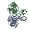
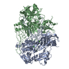
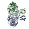
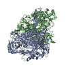
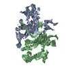
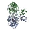
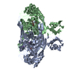
Entry Database : PDB / ID : 6ezzTitle Crystal structure of Escherichia coli amine oxidase mutant E573Q Primary amine oxidase Keywords Function / homology Function Domain/homology Component
/ / / / / / / / / / / / / / / / / / / / / / / / / / / / / / / / / / / / / / / / / / / Biological species Escherichia coli (E. coli)Method / / / Resolution : 1.8 Å Authors Gaule, T.G. / Smith, M.A. / Tych, K.M. / Pirrat, P. / Trinh, C.H. / Pearson, A.R. / Knowles, P.F. / McPherson, M.J. Funding support Organization Grant number Country Biotechnology and Biological Sciences Research Council BB/C00468X/1
Journal : Biochemistry / Year : 2018Title : Oxygen Activation Switch in the Copper Amine Oxidase of Escherichia coli.Authors : Gaule, T.G. / Smith, M.A. / Tych, K.M. / Pirrat, P. / Trinh, C.H. / Pearson, A.R. / Knowles, P.F. / McPherson, M.J. History Deposition Nov 16, 2017 Deposition site / Processing site Revision 1.0 Aug 29, 2018 Provider / Type Revision 1.1 Sep 19, 2018 Group / Database references / Category / citation_authorItem _citation.journal_volume / _citation.page_first ... _citation.journal_volume / _citation.page_first / _citation.page_last / _citation.title / _citation_author.identifier_ORCID Revision 1.2 Jan 17, 2024 Group Data collection / Database references ... Data collection / Database references / Derived calculations / Refinement description Category chem_comp_atom / chem_comp_bond ... chem_comp_atom / chem_comp_bond / database_2 / pdbx_initial_refinement_model / struct_conn Item _database_2.pdbx_DOI / _database_2.pdbx_database_accession ... _database_2.pdbx_DOI / _database_2.pdbx_database_accession / _struct_conn.pdbx_dist_value / _struct_conn.ptnr1_auth_asym_id / _struct_conn.ptnr1_auth_comp_id / _struct_conn.ptnr1_auth_seq_id / _struct_conn.ptnr1_label_asym_id / _struct_conn.ptnr1_label_atom_id / _struct_conn.ptnr1_label_comp_id / _struct_conn.ptnr1_label_seq_id / _struct_conn.ptnr2_auth_asym_id / _struct_conn.ptnr2_auth_comp_id / _struct_conn.ptnr2_auth_seq_id / _struct_conn.ptnr2_label_asym_id / _struct_conn.ptnr2_label_atom_id / _struct_conn.ptnr2_label_comp_id
Show all Show less
 Movie
Movie Controller


 Open data
 Basic information
 Components
 Keywords
 Function and homology information
 X-RAY DIFFRACTION /
  Authors
 United Kingdom, 1items
  Citation
 Structure visualization
 Downloads & links
 Other downloads
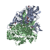


 PDBj
PDBj Assembly

 Mass: 63.546 Da / Num. of mol.: 2 / Source method: obtained synthetically / Formula: Cu
 Mass: 40.078 Da / Num. of mol.: 2 / Source method: obtained synthetically / Formula: Ca
 Mass: 92.094 Da / Num. of mol.: 9 / Source method: obtained synthetically / Formula: C3H8O3
 Mass: 18.015 Da / Num. of mol.: 1113 / Source method: isolated from a natural source / Formula: H2O
 Sample preparation
 Processing