 Movie
Movie Controller

[English] 日本語
 Yorodumi
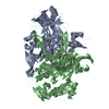
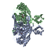
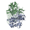
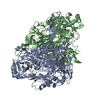
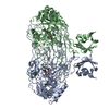
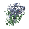
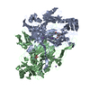
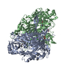
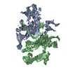
- PDB-1qaf: THE ACTIVE SITE BASE CONTROLS COFACTOR REACTIVITY IN ESCHERICHIA ... -
+ Open data

- Basic information
| Entry | Database: PDB / ID: 1qaf | ||||||
|---|---|---|---|---|---|---|---|
| Title | THE ACTIVE SITE BASE CONTROLS COFACTOR REACTIVITY IN ESCHERICHIA COLI AMINE OXIDASE : X-RAY CRYSTALLOGRAPHIC STUDIES WITH MUTATIONAL VARIANTS | ||||||
 Components Components | PROTEIN (COPPER AMINE OXIDASE) | ||||||
 Keywords Keywords | OXIDOREDUCTASE / CATALYTIC DOMAIN MAINLY ANTI-PARALLEL BETA SHEET / THREE PERIPHERAL ALPHA-BETA DOMAINS | ||||||
| Function / homology |  Function and homology information Function and homology informationphenylethylamine catabolic process / primary-amine oxidase / primary methylamine oxidase activity / amine metabolic process / L-phenylalanine catabolic process / quinone binding / periplasmic space / copper ion binding / calcium ion binding Similarity search - Function | ||||||
| Biological species |  | ||||||
| Method |  X-RAY DIFFRACTION / SYNCHROTRON / DIFFERENCE FOURIER / Resolution: 2.2 Å X-RAY DIFFRACTION / SYNCHROTRON / DIFFERENCE FOURIER / Resolution: 2.2 Å | ||||||
 Authors Authors | Murray, J.M. / Wilmot, C.M. / Saysell, C.G. / Jaeger, J. / Knowles, P.F. / Phillips, S.E. / McPherson, M.J. | ||||||
 Citation Citation | Journal: Biochemistry / Year: 1999 Title: The active site base controls cofactor reactivity in Escherichia coli amine oxidase: x-ray crystallographic studies with mutational variants. Authors: Murray, J.M. / Saysell, C.G. / Wilmot, C.M. / Tambyrajah, W.S. / Jaeger, J. / Knowles, P.F. / Phillips, S.E. / McPherson, M.J. | ||||||
| History |
|
- Structure visualization
| Structure viewer | Molecule: MolmilJmol/JSmol |
|---|
- Downloads & links
-Download
| PDBx/mmCIF format | 1qaf.cif.gz | 330.4 KB | Display | PDBx/mmCIF format |
|---|---|---|---|---|
| PDB format | pdb1qaf.ent.gz | 260.4 KB | Display | PDB format |
| PDBx/mmJSON format | 1qaf.json.gz | Tree view | PDBx/mmJSON format | |
| Others |  Other downloads Other downloads |
-Validation report
| Arichive directory | https://data.pdbj.org/pub/pdb/validation_reports/qa/1qafftp://data.pdbj.org/pub/pdb/validation_reports/qa/1qaf | HTTPS FTP |
|---|
-Related structure data
| Related structure data | 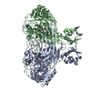 1dyuC  1qakC 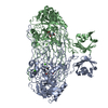 1qalC  1oacS S: Starting model for refinement C: citing same article ( |
|---|---|
| Similar structure data |


-Links
 PDBj
PDBj- Assembly
| Deposited unit | 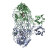
| ||||||||
|---|---|---|---|---|---|---|---|---|---|
| 1 |
| ||||||||
| Unit cell |
|
-Components
| #1: Protein | Mass: 80799.125 Da / Num. of mol.: 2 / Mutation: D383E Source method: isolated from a genetically manipulated source Source: (gene. exp.) #2: Chemical |   Mass: 63.546 Da / Num. of mol.: 2 / Source method: obtained synthetically / Formula: Cu Mass: 63.546 Da / Num. of mol.: 2 / Source method: obtained synthetically / Formula: Cu#3: Chemical | ChemComp-CA /   Mass: 40.078 Da / Num. of mol.: 4 / Source method: obtained synthetically / Formula: Ca Mass: 40.078 Da / Num. of mol.: 4 / Source method: obtained synthetically / Formula: Ca#4: Chemical | ChemComp-GOL / |   Mass: 92.094 Da / Num. of mol.: 1 / Source method: obtained synthetically / Formula: C3H8O3 Mass: 92.094 Da / Num. of mol.: 1 / Source method: obtained synthetically / Formula: C3H8O3#5: Water | ChemComp-HOH / |  Mass: 18.015 Da / Num. of mol.: 1407 / Source method: isolated from a natural source / Formula: H2O Mass: 18.015 Da / Num. of mol.: 1407 / Source method: isolated from a natural source / Formula: H2O |
|---|
-Experimental details
-Experiment
| Experiment | Method: X-RAY DIFFRACTION / Number of used crystals: 1 |
|---|
- Sample preparation
| Crystal | Density Matthews: 2.79 Å3/Da / Density % sol: 55.92 % | |||||||||||||||
|---|---|---|---|---|---|---|---|---|---|---|---|---|---|---|---|---|
| Crystal grow | Temperature: 315 K / Method: vapor diffusion, sitting drop / pH: 7.1 Details: VAPOR DIFFUSION, SITTING DROP PH 7.1, 315 K SODIUM CITRATE, HEPES | |||||||||||||||
| Crystal | *PLUS | |||||||||||||||
| Crystal grow | *PLUS | |||||||||||||||
| Components of the solutions | *PLUS
|
-Data collection
| Diffraction | Mean temperature: 100 K |
|---|---|
| Diffraction source | Source: SYNCHROTRON / Site: SRS  / Beamline: PX7.2 / Wavelength: 1.488 / Beamline: PX7.2 / Wavelength: 1.488 |
| Detector | Type: MARRESEARCH / Detector: IMAGE PLATE / Date: Jun 21, 1997 / Details: MIRRORS |
| Radiation | Protocol: SINGLE WAVELENGTH / Monochromatic (M) / Laue (L): M / Scattering type: x-ray |
| Radiation wavelength | Wavelength: 1.488 Å / Relative weight: 1 |
| Reflection | Resolution: 2.2→20 Å / Num. all: 92396 / Num. obs: 92396 / % possible obs: 91.6 % / Observed criterion σ(F): 1 / Observed criterion σ(I): 1 / Redundancy: 2.9 % / Biso Wilson estimate: 30.2 Å2 / Rmerge(I) obs: 0.066 / Net I/σ(I): 8.3 |
| Reflection shell | Resolution: 2.2→2.3 Å / Redundancy: 1.03 % / Rmerge(I) obs: 0.204 / Mean I/σ(I) obs: 3.2 / % possible all: 73.3 |
| Reflection | *PLUS Num. obs: 84390 |
- Processing
| Software |
| ||||||||||||||||||||||||||||||||||||||||||||||||||||||||||||||||||||||||||||||||||||
|---|---|---|---|---|---|---|---|---|---|---|---|---|---|---|---|---|---|---|---|---|---|---|---|---|---|---|---|---|---|---|---|---|---|---|---|---|---|---|---|---|---|---|---|---|---|---|---|---|---|---|---|---|---|---|---|---|---|---|---|---|---|---|---|---|---|---|---|---|---|---|---|---|---|---|---|---|---|---|---|---|---|---|---|---|---|
| Refinement | Method to determine structure: DIFFERENCE FOURIER Starting model: PDB ENTRY 1OAC Resolution: 2.2→20 Å / SU B: 6.16638 / SU ML: 0.15259 / σ(F): 0 / σ(I): 0 / ESU R: 0.27486 / ESU R Free: 0.22699 / Stereochemistry target values: ENGH & HUBER / Details: USED SIGMA A WEIGHTED MAXIMUM LIKELIHOOD TARGETS.
| ||||||||||||||||||||||||||||||||||||||||||||||||||||||||||||||||||||||||||||||||||||
| Displacement parameters | Biso mean: 32.227 Å2 | ||||||||||||||||||||||||||||||||||||||||||||||||||||||||||||||||||||||||||||||||||||
| Refinement step | Cycle: LAST / Resolution: 2.2→20 Å
| ||||||||||||||||||||||||||||||||||||||||||||||||||||||||||||||||||||||||||||||||||||
| Refine LS restraints |
| ||||||||||||||||||||||||||||||||||||||||||||||||||||||||||||||||||||||||||||||||||||
| Software | *PLUS Name: REFMAC / Classification: refinement | ||||||||||||||||||||||||||||||||||||||||||||||||||||||||||||||||||||||||||||||||||||
| Refinement | *PLUS Highest resolution: 2.2 Å / Lowest resolution: 20 Å / Num. reflection all: 92396 / Num. reflection obs: 82076 / % reflection Rfree: 3.5 % | ||||||||||||||||||||||||||||||||||||||||||||||||||||||||||||||||||||||||||||||||||||
| Solvent computation | *PLUS | ||||||||||||||||||||||||||||||||||||||||||||||||||||||||||||||||||||||||||||||||||||
| Displacement parameters | *PLUS |
