[English] 日本語
Yorodumi
- PDB-2w0q: E. coli copper amine oxidase in complex with Xenon -

+
Open data


ID or keywords:

Loading...

-
Basic information

Entry
Database: PDB / ID: 2w0q
TitleE. coli copper amine oxidase in complex with Xenon
ComponentsCOPPER AMINE OXIDASE
KeywordsOXIDOREDUCTASE / TPQ / XENON / COPPER / E. COLI / CALCIUM / PERIPLASM / OXYGEN ENTRY / METAL-BINDING / COPPER AMINE OXIDASE
Function / homology
Function and homology information


phenylethylamine catabolic process / primary amine oxidase activity / : / : / : / primary-amine oxidase / L-phenylalanine catabolic process / amine metabolic process / quinone binding / periplasmic space ...phenylethylamine catabolic process / primary amine oxidase activity / : / : / : / primary-amine oxidase / L-phenylalanine catabolic process / amine metabolic process / quinone binding / periplasmic space / copper ion binding / calcium ion binding
Similarity search - Function
Copper amine oxidase-like, N-terminal domain / Copper amine oxidase-like, N-terminal / Copper amine oxidase-like, N-terminal domain superfamily / Copper amine oxidase N-terminal domain / Copper Amine Oxidase; Chain A, domain 1 / Copper amine oxidase, N3-terminal / Copper amine oxidase, N2-terminal / Copper amine oxidase, N2 domain / Copper amine oxidase, N3 domain / Copper amine oxidase, catalytic domain ...Copper amine oxidase-like, N-terminal domain / Copper amine oxidase-like, N-terminal / Copper amine oxidase-like, N-terminal domain superfamily / Copper amine oxidase N-terminal domain / Copper Amine Oxidase; Chain A, domain 1 / Copper amine oxidase, N3-terminal / Copper amine oxidase, N2-terminal / Copper amine oxidase, N2 domain / Copper amine oxidase, N3 domain / Copper amine oxidase, catalytic domain / Copper amine oxidase copper-binding site signature. / Copper amine oxidase topaquinone signature. / Nuclear Transport Factor 2; Chain: A, - #40 / Copper amine oxidase / Copper amine oxidase, catalytic domain / Copper amine oxidase, N-terminal / Copper amine oxidase, catalytic domain superfamily / Copper amine oxidase, enzyme domain / Beta-galactosidase; Chain A, domain 5 / Nuclear Transport Factor 2; Chain: A, / Distorted Sandwich / Roll / 2-Layer Sandwich / Mainly Beta / Alpha Beta
Similarity search - Domain/homology
COPPER (II) ION / XENON / Primary amine oxidase
Similarity search - Component
Biological speciesESCHERICHIA COLI (E. coli)
MethodX-RAY DIFFRACTION / SYNCHROTRON / OTHER / Resolution: 2.48 Å
AuthorsPirrat, P. / Smith, M.A. / Pearson, A.R. / McPherson, M.J. / Phillips, S.E.V.
CitationJournal: Acta Crystallogr.,Sect.F / Year: 2008
Title: Structure of a Xenon Derivative of Escherichia Coli Copper Amine Oxidase: Confirmation of the Proposed Oxygen-Entry Pathway.
Authors: Pirrat, P. / Smith, M.A. / Pearson, A.R. / McPherson, M.J. / Phillips, S.E.V.
History
DepositionAug 20, 2008Deposition site: PDBE / Processing site: PDBE
Revision 1.0Dec 16, 2008Provider: repository / Type: Initial release
Revision 1.1May 8, 2011Group: Version format compliance
Revision 1.2Jul 13, 2011Group: Version format compliance
Remark 700 
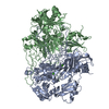
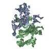
SHEET THE SHEET STRUCTURE OF THIS MOLECULE IS BIFURCATED. IN ORDER TO REPRESENT THIS FEATURE IN ... SHEET THE SHEET STRUCTURE OF THIS MOLECULE IS BIFURCATED. IN ORDER TO REPRESENT THIS FEATURE IN THE SHEET RECORDS BELOW, TWO SHEETS ARE DEFINED.

-
Structure visualization

Structure viewerMolecule:
MolmilJmol/JSmol

Downloads & links

-
Assembly

Deposited unit
A: COPPER AMINE OXIDASE
B: COPPER AMINE OXIDASE
hetero molecules


Theoretical massNumber of molelcules
Total (without water)164,46519
Polymers162,7332
Non-polymers1,73217
Water10,485582
1


  • Idetical with deposited unit
  • defined by author&software
TypeNameSymmetry operationNumber
identity operation1_555x,y,z1
Buried area15620 Å2
ΔGint-94.4 kcal/mol
Surface area62120 Å2
MethodPQS
Unit cell
Length a, b, c (Å)134.588, 166.871, 80.330
Angle α, β, γ (deg.)90.00, 90.00, 90.00
Int Tables number19
Space group name H-MP212121

-
Components

#1: Protein COPPER AMINE OXIDASE / TYRAMINE OXIDASE / 2-PHENYLETHYLAMINE OXIDASE


Mass: 81366.727 Da / Num. of mol.: 2
Source method: isolated from a genetically manipulated source
Source: (gene. exp.) ESCHERICHIA COLI (E. coli) / Plasmid: PKK233-3 / Production host: ESCHERICHIA COLI (E. coli) / Strain (production host): JM109 / References: UniProt: P46883, monoamine oxidase
#2: Chemical ChemComp-CU / COPPER (II) ION / Copper


Mass: 63.546 Da / Num. of mol.: 2 / Source method: obtained synthetically / Formula: Cu
#3: Chemical
ChemComp-CA / CALCIUM ION


Mass: 40.078 Da / Num. of mol.: 4 / Source method: obtained synthetically / Formula: Ca
#4: Chemical
ChemComp-XE / XENON / Xenon


Mass: 131.293 Da / Num. of mol.: 11 / Source method: obtained synthetically / Formula: Xe
#5: Water ChemComp-HOH / water / Water


Mass: 18.015 Da / Num. of mol.: 582 / Source method: isolated from a natural source / Formula: H2O
Nonpolymer detailsEACH MONOMER CONTAINS ONE COPPER ATOM, TWO CALCIUM ATOMS, AND ONE PROTEIN DERIVED COFACTOR, 2,4,5,- ...EACH MONOMER CONTAINS ONE COPPER ATOM, TWO CALCIUM ATOMS, AND ONE PROTEIN DERIVED COFACTOR, 2,4,5,- TRIHYDROXYPHENYL ALANINE QUINONE COFACTOR (TPQ) GENERATED FROM POSTTRANSLATIONAL MODIFICATION OF Y466.

-
Experimental details

-
Experiment

ExperimentMethod: X-RAY DIFFRACTION / Number of used crystals: 1

-
Sample preparation

CrystalDensity Matthews: 2.77 Å3/Da / Density % sol: 55 % / Description: NONE
Crystal growpH: 7.1 / Details: 100MM HEPES, PH 7.1, 1.2 M SODIUM CITRATE

-
Data collection

DiffractionMean temperature: 100 K
Diffraction sourceSource: SYNCHROTRON / Site: SRS / Beamline: PX14.1 / Wavelength: 1.48
DetectorType: ADSC CCD / Detector: CCD
RadiationProtocol: SINGLE WAVELENGTH / Monochromatic (M) / Laue (L): M / Scattering type: x-ray
Radiation wavelengthWavelength: 1.48 Å / Relative weight: 1
ReflectionResolution: 2.48→49.27 Å / Num. obs: 60358 / % possible obs: 98.1 % / Redundancy: 10.1 % / Rmerge(I) obs: 0.12 / Net I/σ(I): 23.5
Reflection shellResolution: 2.48→2.54 Å / Redundancy: 7.4 % / Rmerge(I) obs: 0.45 / Mean I/σ(I) obs: 4.4 / % possible all: 87.3

-
Processing

Software
NameVersionClassification
REFMAC5.2.0019refinement
MOSFLMdata reduction
SCALAdata scaling
RefinementMethod to determine structure: OTHER
Starting model: NONE

Resolution: 2.48→49.27 Å / Cor.coef. Fo:Fc: 0.964 / Cor.coef. Fo:Fc free: 0.931 / SU B: 6.732 / SU ML: 0.154 / Cross valid method: THROUGHOUT / ESU R: 0.392 / ESU R Free: 0.247 / Stereochemistry target values: MAXIMUM LIKELIHOOD / Details: HYDROGENS HAVE BEEN ADDED IN THE RIDING POSITIONS.
RfactorNum. reflection% reflectionSelection details
Rfree0.219 3207 5 %RANDOM
Rwork0.158 ---
obs0.161 60358 98.1 %-
Solvent computationIon probe radii: 0.8 Å / Shrinkage radii: 0.8 Å / VDW probe radii: 1.2 Å / Solvent model: MASK
Displacement parametersBiso mean: 40.67 Å2
Baniso -1Baniso -2Baniso -3
1-0.14 Å20 Å20 Å2
2--0.15 Å20 Å2
3----0.29 Å2
Refinement stepCycle: LAST / Resolution: 2.48→49.27 Å
ProteinNucleic acidLigandSolventTotal
Num. atoms11348 0 17 582 11947
Refine LS restraints
Refine-IDTypeDev idealDev ideal targetNumber
X-RAY DIFFRACTIONr_bond_refined_d0.0170.02211633
X-RAY DIFFRACTIONr_bond_other_d
X-RAY DIFFRACTIONr_angle_refined_deg1.6511.94915833
X-RAY DIFFRACTIONr_angle_other_deg
X-RAY DIFFRACTIONr_dihedral_angle_1_deg6.69251431
X-RAY DIFFRACTIONr_dihedral_angle_2_deg37.04724.936543
X-RAY DIFFRACTIONr_dihedral_angle_3_deg16.917151914
X-RAY DIFFRACTIONr_dihedral_angle_4_deg18.6931554
X-RAY DIFFRACTIONr_chiral_restr0.1060.21725
X-RAY DIFFRACTIONr_gen_planes_refined0.0060.028952
X-RAY DIFFRACTIONr_gen_planes_other
X-RAY DIFFRACTIONr_nbd_refined0.2320.25033
X-RAY DIFFRACTIONr_nbd_other
X-RAY DIFFRACTIONr_nbtor_refined0.3180.27839
X-RAY DIFFRACTIONr_nbtor_other
X-RAY DIFFRACTIONr_xyhbond_nbd_refined0.1620.2737
X-RAY DIFFRACTIONr_xyhbond_nbd_other
X-RAY DIFFRACTIONr_metal_ion_refined
X-RAY DIFFRACTIONr_metal_ion_other
X-RAY DIFFRACTIONr_symmetry_vdw_refined0.2120.240
X-RAY DIFFRACTIONr_symmetry_vdw_other
X-RAY DIFFRACTIONr_symmetry_hbond_refined0.1430.25
X-RAY DIFFRACTIONr_symmetry_hbond_other
X-RAY DIFFRACTIONr_symmetry_metal_ion_refined
X-RAY DIFFRACTIONr_symmetry_metal_ion_other
X-RAY DIFFRACTIONr_mcbond_it0.8781.57167
X-RAY DIFFRACTIONr_mcbond_other
X-RAY DIFFRACTIONr_mcangle_it1.69211633
X-RAY DIFFRACTIONr_mcangle_other
X-RAY DIFFRACTIONr_scbond_it2.73634728
X-RAY DIFFRACTIONr_scbond_other
X-RAY DIFFRACTIONr_scangle_it4.5364.54200
X-RAY DIFFRACTIONr_scangle_other
X-RAY DIFFRACTIONr_long_range_B_refined
X-RAY DIFFRACTIONr_long_range_B_other
X-RAY DIFFRACTIONr_rigid_bond_restr
X-RAY DIFFRACTIONr_sphericity_free
X-RAY DIFFRACTIONr_sphericity_bonded
LS refinement shellResolution: 2.48→2.54 Å / Total num. of bins used: 20 /
RfactorNum. reflection
Rfree0.301 220
Rwork0.219 3900

+
About Yorodumi

-
News

-
Feb 9, 2022. New format data for meta-information of EMDB entries

New format data for meta-information of EMDB entries

  • Version 3 of the EMDB header file is now the official format.
  • The previous official version 1.9 will be removed from the archive.

Related info.:EMDB header

External links:wwPDB to switch to version 3 of the EMDB data model

-
Aug 12, 2020. Covid-19 info

Covid-19 info

URL: https://pdbj.org/emnavi/covid19.php

New page: Covid-19 featured information page in EM Navigator.

Related info.:Covid-19 info / Mar 5, 2020. Novel coronavirus structure data

+
Mar 5, 2020. Novel coronavirus structure data

Novel coronavirus structure data

Related info.:Yorodumi Speices / Aug 12, 2020. Covid-19 info

External links:COVID-19 featured content - PDBj / Molecule of the Month (242):Coronavirus Proteases

+
Jan 31, 2019. EMDB accession codes are about to change! (news from PDBe EMDB page)

EMDB accession codes are about to change! (news from PDBe EMDB page)

  • The allocation of 4 digits for EMDB accession codes will soon come to an end. Whilst these codes will remain in use, new EMDB accession codes will include an additional digit and will expand incrementally as the available range of codes is exhausted. The current 4-digit format prefixed with “EMD-” (i.e. EMD-XXXX) will advance to a 5-digit format (i.e. EMD-XXXXX), and so on. It is currently estimated that the 4-digit codes will be depleted around Spring 2019, at which point the 5-digit format will come into force.
  • The EM Navigator/Yorodumi systems omit the EMD- prefix.

Related info.:Q: What is EMD? / ID/Accession-code notation in Yorodumi/EM Navigator

External links:EMDB Accession Codes are Changing Soon! / Contact to PDBj

+
Jul 12, 2017. Major update of PDB

Major update of PDB

  • wwPDB released updated PDB data conforming to the new PDBx/mmCIF dictionary.
  • This is a major update changing the version number from 4 to 5, and with Remediation, in which all the entries are updated.
  • In this update, many items about electron microscopy experimental information are reorganized (e.g. em_software).
  • Now, EM Navigator and Yorodumi are based on the updated data.

External links:wwPDB Remediation / Enriched Model Files Conforming to OneDep Data Standards Now Available in the PDB FTP Archive

-
Yorodumi

Thousand views of thousand structures

  • Yorodumi is a browser for structure data from EMDB, PDB, SASBDB, etc.
  • This page is also the successor to EM Navigator detail page, and also detail information page/front-end page for Omokage search.
  • The word "yorodu" (or yorozu) is an old Japanese word meaning "ten thousand". "mi" (miru) is to see.

Related info.:EMDB / PDB / SASBDB / Comparison of 3 databanks / Yorodumi Search / Aug 31, 2016. New EM Navigator & Yorodumi / Yorodumi Papers / Jmol/JSmol / Function and homology information / Changes in new EM Navigator and Yorodumi

Read more