 Movie
Movie Controller

[English] 日本語
 Yorodumi
- PDB-1dyu: The active site base controls cofactor reactivity in Escherichia ... -
+ Open data

- Basic information
| Entry | Database: PDB / ID: 1dyu | ||||||
|---|---|---|---|---|---|---|---|
| Title | The active site base controls cofactor reactivity in Escherichia coli amine oxidase: X-ray crystallographic studies with mutational variants. | ||||||
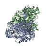
 Components Components | COPPER AMINE OXIDASE | ||||||
 Keywords Keywords | OXIDOREDUCTASE / TPQ / COPPER AMINE OXIDASE / EC 1.4.3.4 / WILDTYPE / CATALYTIC BASE | ||||||
| Function / homology |  Function and homology information Function and homology informationphenylethylamine catabolic process / primary-amine oxidase / primary methylamine oxidase activity / amine metabolic process / L-phenylalanine catabolic process / quinone binding / periplasmic space / copper ion binding / calcium ion binding Similarity search - Function | ||||||
| Biological species |  | ||||||
| Method |  X-RAY DIFFRACTION / SYNCHROTRON / MOLECULAR REPLACEMENT / Resolution: 2.04 Å X-RAY DIFFRACTION / SYNCHROTRON / MOLECULAR REPLACEMENT / Resolution: 2.04 Å | ||||||
 Authors Authors | Murray, J.M. / Wilmot, C.M. / Saysell, C.G. / Jaeger, J. / Knowles, P.F. / Phillips, S.E.V. / McPherson, M.J. | ||||||
 Citation Citation | Journal: Biochemistry / Year: 1999 Title: The Active Site Base Controls Cofactor Reactivity in Escherichia Coli Amine Oxidase : X-Ray Crystallographicstudies with Mutational Variants Authors: Murray, J.M. / Wilmot, C.M. / Saysell, C.G. / Jaeger, J. / Knowles, P.F. / Phillips, S.E.V. / McPherson, M.J. | ||||||
| History |
| ||||||
| Remark 700 | SHEET DETERMINATION METHOD: DSSP THERE ARE SEVERAL BIFURCATED SHEETS IN THIS STRUCTURE. EACH IS ... SHEET DETERMINATION METHOD: DSSP THERE ARE SEVERAL BIFURCATED SHEETS IN THIS STRUCTURE. EACH IS REPRESENTED BY TWO SHEETS WHICH HAVE ONE OR MORE IDENTICAL STRANDS. SHEETS C AND C1 REPRESENT ONE BIFURCATED SHEET. SHEETS K AND K1 REPRESENT ONE BIFURCATED SHEET. |
- Structure visualization
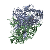
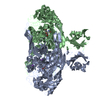
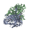
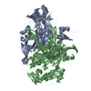
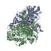
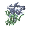
| Structure viewer | Molecule: MolmilJmol/JSmol |
|---|
- Downloads & links
-Download
| PDBx/mmCIF format | 1dyu.cif.gz | 326.3 KB | Display | PDBx/mmCIF format |
|---|---|---|---|---|
| PDB format | pdb1dyu.ent.gz | 263 KB | Display | PDB format |
| PDBx/mmJSON format | 1dyu.json.gz | Tree view | PDBx/mmJSON format | |
| Others |  Other downloads Other downloads |
-Validation report
| Arichive directory | https://data.pdbj.org/pub/pdb/validation_reports/dy/1dyuftp://data.pdbj.org/pub/pdb/validation_reports/dy/1dyu | HTTPS FTP |
|---|
-Related structure data
| Related structure data | 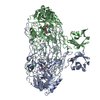 1qafC  1qakC 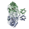 1qalC  1oacS S: Starting model for refinement C: citing same article ( |
|---|---|
| Similar structure data |


-Links
 PDBj
PDBj- Assembly
| Deposited unit | 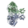
| ||||||||
|---|---|---|---|---|---|---|---|---|---|
| 1 |
| ||||||||
| Unit cell |
| ||||||||
| Details | BIOLOGICAL_UNIT: DIMERIC |
-Components
| #1: Protein | Mass: 81366.727 Da / Num. of mol.: 2 Source method: isolated from a genetically manipulated source Details: EACH MONOMER CONTAINS ONE COPPER ATOM, TWO CALCIUM ATOMS, AND ONE 2,4,5, -TRIHYDROXYPHENYLALANINE QUINONE COFACTOR. Source: (gene. exp.) Description: CLONED AND OVER-EXPRESSED IN E. COLI USING PLASMID PKK233-3 Cellular location: PERIPLASM / Plasmid: PLASMID DERIVED FROM PKK233-3 / Cellular location (production host): PERIPLASM / Production host: #2: Chemical |   Mass: 63.546 Da / Num. of mol.: 2 / Source method: obtained synthetically / Formula: Cu Mass: 63.546 Da / Num. of mol.: 2 / Source method: obtained synthetically / Formula: Cu#3: Chemical | ChemComp-CA /   Mass: 40.078 Da / Num. of mol.: 4 / Source method: obtained synthetically / Formula: Ca Mass: 40.078 Da / Num. of mol.: 4 / Source method: obtained synthetically / Formula: Ca#4: Water | ChemComp-HOH / |  Mass: 18.015 Da / Num. of mol.: 1568 / Source method: isolated from a natural source / Formula: H2O Mass: 18.015 Da / Num. of mol.: 1568 / Source method: isolated from a natural source / Formula: H2O |
|---|
-Experimental details
-Experiment
| Experiment | Method: X-RAY DIFFRACTION / Number of used crystals: 1 |
|---|
- Sample preparation
| Crystal | Density Matthews: 2.73 Å3/Da / Density % sol: 55 % | |||||||||||||||
|---|---|---|---|---|---|---|---|---|---|---|---|---|---|---|---|---|
| Crystal grow | pH: 7.1 / Details: 1.2 M SODIUM CITRATE, 0.1 M HEPES PH 7.1 | |||||||||||||||
| Crystal grow | *PLUS Method: vapor diffusion, sitting drop | |||||||||||||||
| Components of the solutions | *PLUS
|
-Data collection
| Diffraction | Mean temperature: 100 K |
|---|---|
| Diffraction source | Source: SYNCHROTRON / Site: ESRF  / Beamline: ID2 / Wavelength: 0.99 / Beamline: ID2 / Wavelength: 0.99 |
| Detector | Type: MARRESEARCH / Detector: IMAGE PLATE |
| Radiation | Protocol: SINGLE WAVELENGTH / Monochromatic (M) / Laue (L): M / Scattering type: x-ray |
| Radiation wavelength | Wavelength: 0.99 Å / Relative weight: 1 |
| Reflection | Resolution: 2.04→20 Å / Num. obs: 105826 / % possible obs: 93.3 % / Redundancy: 3.2 % / Biso Wilson estimate: 36 Å2 / Rsym value: 0.06 / Net I/σ(I): 7.9 |
| Reflection shell | Resolution: 2.04→2.38 Å / Redundancy: 3.2 % / Rmerge(I) obs: 0.07 / Mean I/σ(I) obs: 3.4 / Rsym value: 0.19 / % possible all: 91 |
| Reflection | *PLUS Rmerge(I) obs: 0.058 |
| Reflection shell | *PLUS % possible obs: 91 % |
- Processing
| Software |
| ||||||||||||||||||||||||||||||||||||||||||||||||||||||||||||||||||||||||||||||||||||
|---|---|---|---|---|---|---|---|---|---|---|---|---|---|---|---|---|---|---|---|---|---|---|---|---|---|---|---|---|---|---|---|---|---|---|---|---|---|---|---|---|---|---|---|---|---|---|---|---|---|---|---|---|---|---|---|---|---|---|---|---|---|---|---|---|---|---|---|---|---|---|---|---|---|---|---|---|---|---|---|---|---|---|---|---|---|
| Refinement | Method to determine structure: MOLECULAR REPLACEMENT Starting model: PDB ENTRY 1OAC Resolution: 2.04→20 Å / Cross valid method: THROUGHOUT / σ(F): 0 Details: NEITHER THE N-TERMINAL OR C-TERMINAL AMINO ACIDS WERE WELL DEFINED BY THE ELECTRON DENSITY
| ||||||||||||||||||||||||||||||||||||||||||||||||||||||||||||||||||||||||||||||||||||
| Displacement parameters | Biso mean: 31.9 Å2
| ||||||||||||||||||||||||||||||||||||||||||||||||||||||||||||||||||||||||||||||||||||
| Refinement step | Cycle: LAST / Resolution: 2.04→20 Å
| ||||||||||||||||||||||||||||||||||||||||||||||||||||||||||||||||||||||||||||||||||||
| Refine LS restraints |
|
