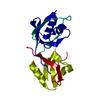
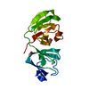
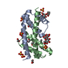
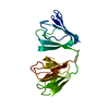
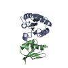
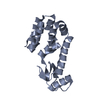
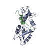
Entry Database : PDB / ID : 1jyqTitle Xray Structure of Grb2 SH2 Domain Complexed with a Highly Affine Phospho Peptide GROWTH FACTOR RECEPTOR-BOUND PROTEIN 2 mAZ-pY-(alpha Me)pY-N-NH2 peptide inhibitor Keywords / / / / Function / homology Function Domain/homology Component
/ / / / / / / / / / / / / / / / / / / / / / / / / / / / / / / / / / / / / / / / / / / / / / / / / / / / / / / / / / / / / / / / / / / / / / / / / / / / / / / / / / / / / / / / / / / / / / / / / / / / / / / / / / / / / / / / / / / / / / / / / / / / / / / / / / / / / Biological species Homo sapiens (human)Method / / / Resolution : 2 Å Authors Nioche, P. / Liu, W.-Q. / Broutin, I. / Charbonnier, F. / Latreille, M.-T. / Vidal, M. / Roques, B. / Garbay, C. / Ducruix, A. Journal : J.Mol.Biol. / Year : 2002Title : Crystal structures of the SH2 domain of Grb2: highlight on the binding of a new high-affinity inhibitor.Authors : Nioche, P. / Liu, W.Q. / Broutin, I. / Charbonnier, F. / Latreille, M.T. / Vidal, M. / Roques, B. / Garbay, C. / Ducruix, A. History Deposition Sep 13, 2001 Deposition site / Processing site Revision 1.0 Mar 13, 2002 Provider / Type Revision 1.1 Apr 27, 2008 Group Revision 1.2 Jul 13, 2011 Group Atomic model / Database references ... Atomic model / Database references / Derived calculations / Non-polymer description / Structure summary / Version format compliance Revision 1.3 Dec 12, 2012 Group Revision 1.4 Apr 4, 2018 Group / Category / Item Revision 2.0 Jul 10, 2024 Group Data collection / Database references ... Data collection / Database references / Derived calculations / Non-polymer description / Structure summary Category chem_comp / chem_comp_atom ... chem_comp / chem_comp_atom / chem_comp_bond / database_2 / entity / struct_conn / struct_ref_seq_dif Item _chem_comp.formula / _chem_comp.formula_weight ... _chem_comp.formula / _chem_comp.formula_weight / _database_2.pdbx_DOI / _database_2.pdbx_database_accession / _entity.formula_weight / _struct_conn.pdbx_dist_value / _struct_conn.pdbx_leaving_atom_flag / _struct_conn.ptnr1_auth_asym_id / _struct_conn.ptnr1_label_asym_id / _struct_conn.ptnr2_auth_asym_id / _struct_conn.ptnr2_label_asym_id / _struct_ref_seq_dif.details Revision 2.1 Nov 20, 2024 Group / Category / pdbx_modification_feature
Show all Show less
 Movie
Movie Controller

 Yorodumi
 Open data
 Basic information
 Components
 Keywords
 Function and homology information
 Homo sapiens (human)
 X-RAY DIFFRACTION /
  Authors
 Citation
 Structure visualization
 Downloads & links
 Other downloads
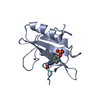

 PDBj
PDBj


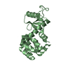
 Assembly
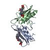


 Type: Peptide-like / Class: Inhibitor / Mass: 781.597 Da / Num. of mol.: 2 / Source method: obtained synthetically / Details: This peptide was chemically synthesized / References: MAZ-PY-(ALPHA ME)PY-N-NH2
 Mass: 18.015 Da / Num. of mol.: 166 / Source method: isolated from a natural source / Formula: H2O
 Sample preparation
 / Type:
  Processing