 Movie
Movie Controller

[English] 日本語
 Yorodumi
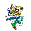
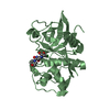
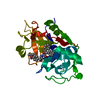
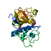
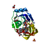
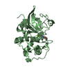
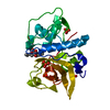
- PDB-1iwd: Proposed Amino Acid Sequence and the 1.63 Angstrom X-ray Crystal ... -
+ Open data

- Basic information
| Entry | Database: PDB / ID: 1iwd | ||||||
|---|---|---|---|---|---|---|---|
| Title | Proposed Amino Acid Sequence and the 1.63 Angstrom X-ray Crystal Structure of a Plant Cysteine Protease Ervatamin B: Insight into the Structural Basis of its Stability and Substrate Specificity. | ||||||
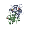
 Components Components | ERVATAMIN B | ||||||
 Keywords Keywords | HYDROLASE / Cysteine protease / alpha-beta protein / catalytic dyad / L-domain / R-domain. | ||||||
| Function / homology |  Function and homology information Function and homology informationcysteine-type peptidase activity / Hydrolases; Acting on peptide bonds (peptidases); Cysteine endopeptidases / proteolysis / extracellular region Similarity search - Function | ||||||
| Biological species |  Tabernaemontana divaricata (pinwheelflower) Tabernaemontana divaricata (pinwheelflower) | ||||||
| Method |  X-RAY DIFFRACTION / MOLECULAR REPLACEMENT / Resolution: 1.63 Å X-RAY DIFFRACTION / MOLECULAR REPLACEMENT / Resolution: 1.63 Å | ||||||
 Authors Authors | Chakrabarti, C. / Biswas, S. / Dattagupta, J.K. | ||||||
 Citation Citation | Journal: Proteins / Year: 2003 Title: Proposed amino acid sequence and the 1.63 A X-ray crystal structure of a plant cysteine protease, ervatamin B: Some insights into the structural basis of its stability and substrate specificity Authors: Biswas, S. / Chakrabarti, C. / Kundu, S. / Jagannadham, M.V. / Dattagupta, J.K. | ||||||
| History |
|
- Structure visualization
| Structure viewer | Molecule: MolmilJmol/JSmol |
|---|
- Downloads & links
-Download
| PDBx/mmCIF format | 1iwd.cif.gz | 57.9 KB | Display | PDBx/mmCIF format |
|---|---|---|---|---|
| PDB format | pdb1iwd.ent.gz | 41.4 KB | Display | PDB format |
| PDBx/mmJSON format | 1iwd.json.gz | Tree view | PDBx/mmJSON format | |
| Others |  Other downloads Other downloads |
-Validation report
| Arichive directory | https://data.pdbj.org/pub/pdb/validation_reports/iw/1iwdftp://data.pdbj.org/pub/pdb/validation_reports/iw/1iwd | HTTPS FTP |
|---|
-Related structure data
| Related structure data | 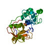 2actS S: Starting model for refinement |
|---|---|
| Similar structure data |
-Links
 PDBj
PDBj
- Assembly
| Deposited unit | 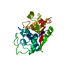
| ||||||||
|---|---|---|---|---|---|---|---|---|---|
| 1 |
| ||||||||
| Unit cell |
|
-Components
| #1: Protein | Mass: 23200.805 Da / Num. of mol.: 1 / Source method: isolated from a natural source Source: (natural) Tabernaemontana divaricata (pinwheelflower)References: UniProt: P60994 |
|---|---|
| #2: Chemical | ChemComp-THJ / 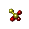  Mass: 112.128 Da / Num. of mol.: 1 / Source method: obtained synthetically / Formula: O3S2 Mass: 112.128 Da / Num. of mol.: 1 / Source method: obtained synthetically / Formula: O3S2 |
| #3: Water | ChemComp-HOH /  Mass: 18.015 Da / Num. of mol.: 208 / Source method: isolated from a natural source / Formula: H2O Mass: 18.015 Da / Num. of mol.: 208 / Source method: isolated from a natural source / Formula: H2O |
| Has protein modification | Y |
-Experimental details
-Experiment
| Experiment | Method: X-RAY DIFFRACTION / Number of used crystals: 1 |
|---|
- Sample preparation
| Crystal | Density Matthews: 1.9 Å3/Da / Density % sol: 34.79 % | |||||||||||||||||||||||||||||||||||
|---|---|---|---|---|---|---|---|---|---|---|---|---|---|---|---|---|---|---|---|---|---|---|---|---|---|---|---|---|---|---|---|---|---|---|---|---|
| Crystal grow | Temperature: 292 K / Method: vapor diffusion, hanging drop / pH: 8.5 Details: PEG4000, Tris HCl, Magnesium Chloride, pH 8.5, VAPOR DIFFUSION, HANGING DROP, temperature 292K | |||||||||||||||||||||||||||||||||||
| Crystal grow | *PLUS | |||||||||||||||||||||||||||||||||||
| Components of the solutions | *PLUS
|
-Data collection
| Diffraction | Mean temperature: 293 K |
|---|---|
| Diffraction source | Source: ROTATING ANODE / Type: RIGAKU RU200 / Wavelength: 1.54 Å |
| Detector | Type: MARRESEARCH / Detector: IMAGE PLATE / Date: Mar 9, 1999 / Details: osmic Max-Flux confocal optics |
| Radiation | Monochromator: Osmic Max-Flux optic system / Protocol: SINGLE WAVELENGTH / Monochromatic (M) / Laue (L): M / Scattering type: x-ray |
| Radiation wavelength | Wavelength: 1.54 Å / Relative weight: 1 |
| Reflection | Resolution: 1.63→50 Å / Num. all: 25294 / Num. obs: 25048 / % possible obs: 99.1 % / Observed criterion σ(F): -3 / Redundancy: 5.6 % / Biso Wilson estimate: 18.1 Å2 / Rmerge(I) obs: 0.048 / Net I/σ(I): 24.06 |
| Reflection shell | Resolution: 1.63→1.67 Å / Rmerge(I) obs: 0.281 / Mean I/σ(I) obs: 4 / % possible all: 89.2 |
| Reflection | *PLUS Lowest resolution: 50 Å / Num. obs: 25294 / Num. measured all: 142404 |
| Reflection shell | *PLUS % possible obs: 89.2 % |
- Processing
| Software |
| ||||||||||||||||||||||||||||||||||||
|---|---|---|---|---|---|---|---|---|---|---|---|---|---|---|---|---|---|---|---|---|---|---|---|---|---|---|---|---|---|---|---|---|---|---|---|---|---|
| Refinement | Method to determine structure: MOLECULAR REPLACEMENT Starting model: PDB entry 2ACT Resolution: 1.63→14.94 Å / Rfactor Rfree error: 0.005 / Isotropic thermal model: RESTRAINED / Cross valid method: THROUGHOUT / σ(F): 0 / Stereochemistry target values: Engh & Huber
| ||||||||||||||||||||||||||||||||||||
| Solvent computation | Solvent model: FLAT MODEL / Bsol: 47.353 Å2 / ksol: 0.356996 e/Å3 | ||||||||||||||||||||||||||||||||||||
| Displacement parameters | Biso mean: 17.8 Å2
| ||||||||||||||||||||||||||||||||||||
| Refine analyze | Luzzati coordinate error free: 0.17 Å / Luzzati sigma a free: 0.12 Å | ||||||||||||||||||||||||||||||||||||
| Refinement step | Cycle: LAST / Resolution: 1.63→14.94 Å
| ||||||||||||||||||||||||||||||||||||
| Refine LS restraints |
| ||||||||||||||||||||||||||||||||||||
| LS refinement shell | Resolution: 1.63→1.73 Å / Rfactor Rfree error: 0.018 / Total num. of bins used: 6
| ||||||||||||||||||||||||||||||||||||
| Xplor file |
| ||||||||||||||||||||||||||||||||||||
| Refinement | *PLUS Lowest resolution: 15 Å / % reflection Rfree: 5 % | ||||||||||||||||||||||||||||||||||||
| Solvent computation | *PLUS | ||||||||||||||||||||||||||||||||||||
| Displacement parameters | *PLUS | ||||||||||||||||||||||||||||||||||||
| Refine LS restraints | *PLUS
| ||||||||||||||||||||||||||||||||||||
| LS refinement shell | *PLUS Rfactor Rfree: 0.1819 / Num. reflection Rfree: 1189 / Rfactor Rwork: 0.1593 / Num. reflection obs: 23348 |
