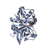
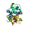
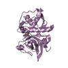
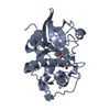
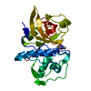
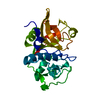
Assembly of collagen fibrils and other multimeric structures / Trafficking and processing of endosomal TLR / cathepsin S / regulation of antigen processing and presentation / Degradation of the extracellular matrix / positive regulation of cation channel activity / basement membrane disassembly / Degradation of CDH1 / sensory perception of itch / antigen processing and presentation of peptide antigen ...Assembly of collagen fibrils and other multimeric structures / Trafficking and processing of endosomal TLR / cathepsin S / regulation of antigen processing and presentation / Degradation of the extracellular matrix / positive regulation of cation channel activity / basement membrane disassembly / Degradation of CDH1 / sensory perception of itch / antigen processing and presentation of peptide antigen / MHC class II antigen presentation / proteoglycan binding / response to acidic pH / collagen catabolic process / fibronectin binding / bone resorption / collagen binding / phagocytic vesicle / Neutrophil degranulation / laminin binding / cysteine-type peptidase activity / early endosome lumen / : / protein processing / antigen processing and presentation of exogenous peptide antigen via MHC class II / positive regulation of inflammatory response / late endosome / peptidase activity / lysosome / cysteine-type endopeptidase activity / cell surface / proteolysis / : / membrane Similarity search - Function
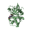
Cathepsin propeptide inhibitor domain (I29) / Cathepsin propeptide inhibitor domain (I29) / Cathepsin propeptide inhibitor domain (I29) / Papain-like cysteine endopeptidase / Cysteine peptidase, asparagine active site / Eukaryotic thiol (cysteine) proteases asparagine active site. / Cysteine peptidase, histidine active site / Eukaryotic thiol (cysteine) proteases histidine active site. / : / Peptidase C1A, papain C-terminal ...Cathepsin propeptide inhibitor domain (I29) / Cathepsin propeptide inhibitor domain (I29) / Cathepsin propeptide inhibitor domain (I29) / Papain-like cysteine endopeptidase / Cysteine peptidase, asparagine active site / Eukaryotic thiol (cysteine) proteases asparagine active site. / Cysteine peptidase, histidine active site / Eukaryotic thiol (cysteine) proteases histidine active site. / : / Peptidase C1A, papain C-terminal / Papain family cysteine protease / Papain family cysteine protease / Cysteine proteinases / Cysteine peptidase, cysteine active site / Eukaryotic thiol (cysteine) proteases cysteine active site. / Cathepsin B; Chain A / Papain-like cysteine peptidase superfamily / Alpha-Beta Complex / Alpha Beta Similarity search - Domain/homology
In the structure databanks used in Yorodumi, some data are registered as the other names, "COVID-19 virus" and "2019-nCoV". Here are the details of the virus and the list of structure data.
Jan 31, 2019. EMDB accession codes are about to change! (news from PDBe EMDB page)
EMDB accession codes are about to change! (news from PDBe EMDB page)
The allocation of 4 digits for EMDB accession codes will soon come to an end. Whilst these codes will remain in use, new EMDB accession codes will include an additional digit and will expand incrementally as the available range of codes is exhausted. The current 4-digit format prefixed with “EMD-” (i.e. EMD-XXXX) will advance to a 5-digit format (i.e. EMD-XXXXX), and so on. It is currently estimated that the 4-digit codes will be depleted around Spring 2019, at which point the 5-digit format will come into force.
The EM Navigator/Yorodumi systems omit the EMD- prefix.
Related info.:Q: What is EMD? / ID/Accession-code notation in Yorodumi/EM Navigator
Yorodumi is a browser for structure data from EMDB, PDB, SASBDB, etc.
This page is also the successor to EM Navigator detail page, and also detail information page/front-end page for Omokage search.
The word "yorodu" (or yorozu) is an old Japanese word meaning "ten thousand". "mi" (miru) is to see.
Related info.:EMDB / PDB / SASBDB / Comparison of 3 databanks / Yorodumi Search / Aug 31, 2016. New EM Navigator & Yorodumi / Yorodumi Papers / Jmol/JSmol / Function and homology information / Changes in new EM Navigator and Yorodumi
 Movie
Movie Controller

 Yorodumi
 Open data
 Basic information
 Components
 Keywords
 Function and homology information
 X-RAY DIFFRACTION /
  Authors
 Citation
 Structure visualization
 Downloads & links
 Other downloads


 PDBj
PDBj


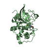
 Assembly
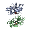


 Mass: 442.917 Da / Num. of mol.: 2 / Source method: obtained synthetically / Formula: C18H23ClN4O5S
 Mass: 18.015 Da / Num. of mol.: 275 / Source method: isolated from a natural source / Formula: H2O
 Sample preparation
 / Beamline: X10SA / Wavelength: 1 Å
 Processing