[English] 日本語
Yorodumi
- PDB-1glo: Crystal Structure of Cys25Ser mutant of human cathepsin S -

+
Open data


ID or keywords:

Loading...

-
Basic information

Entry
Database: PDB / ID: 1glo
TitleCrystal Structure of Cys25Ser mutant of human cathepsin S
ComponentsCATHEPSIN S
KeywordsHYDROLASE / CATHEPSIN S / PROTEINASE / INHIBITOR / THIOL PROTEAS
Function / homology
Function and homology information


cathepsin S / basement membrane disassembly / positive regulation of cation channel activity / antigen processing and presentation of peptide antigen / endolysosome lumen / response to acidic pH / cellular response to thyroid hormone stimulus / Trafficking and processing of endosomal TLR / proteoglycan binding / Assembly of collagen fibrils and other multimeric structures ...cathepsin S / basement membrane disassembly / positive regulation of cation channel activity / antigen processing and presentation of peptide antigen / endolysosome lumen / response to acidic pH / cellular response to thyroid hormone stimulus / Trafficking and processing of endosomal TLR / proteoglycan binding / Assembly of collagen fibrils and other multimeric structures / toll-like receptor signaling pathway / antigen processing and presentation / cysteine-type endopeptidase activator activity involved in apoptotic process / fibronectin binding / collagen catabolic process / extracellular matrix disassembly / laminin binding / collagen binding / MHC class II antigen presentation / phagocytic vesicle / Degradation of the extracellular matrix / lysosomal lumen / proteolysis involved in protein catabolic process / Endosomal/Vacuolar pathway / positive regulation of apoptotic signaling pathway / protein processing / antigen processing and presentation of exogenous peptide antigen via MHC class II / late endosome / tertiary granule lumen / collagen-containing extracellular matrix / adaptive immune response / ficolin-1-rich granule lumen / lysosome / immune response / cysteine-type endopeptidase activity / intracellular membrane-bounded organelle / serine-type endopeptidase activity / Neutrophil degranulation / proteolysis / extracellular space / extracellular region
Similarity search - Function
Cathepsin propeptide inhibitor domain (I29) / Cathepsin propeptide inhibitor domain (I29) / Cathepsin propeptide inhibitor domain (I29) / Papain-like cysteine endopeptidase / : / Cysteine peptidase, asparagine active site / Eukaryotic thiol (cysteine) proteases asparagine active site. / Cysteine peptidase, histidine active site / Eukaryotic thiol (cysteine) proteases histidine active site. / Peptidase C1A, papain C-terminal ...Cathepsin propeptide inhibitor domain (I29) / Cathepsin propeptide inhibitor domain (I29) / Cathepsin propeptide inhibitor domain (I29) / Papain-like cysteine endopeptidase / : / Cysteine peptidase, asparagine active site / Eukaryotic thiol (cysteine) proteases asparagine active site. / Cysteine peptidase, histidine active site / Eukaryotic thiol (cysteine) proteases histidine active site. / Peptidase C1A, papain C-terminal / Papain family cysteine protease / Papain family cysteine protease / Cysteine proteinases / Cysteine peptidase, cysteine active site / Eukaryotic thiol (cysteine) proteases cysteine active site. / Cathepsin B; Chain A / Papain-like cysteine peptidase superfamily / Alpha-Beta Complex / Alpha Beta
Similarity search - Domain/homology
Biological speciesHOMO SAPIENS (human)
MethodX-RAY DIFFRACTION / MOLECULAR REPLACEMENT / Resolution: 2.2 Å
AuthorsTurkenburg, J.P. / Lamers, M.B.A.C. / Brzozowski, A.M. / Wright, L.M. / Hubbard, R.E. / Sturt, S.L. / Williams, D.H.
CitationJournal: Acta Crystallogr.,Sect.D / Year: 2002
Title: Structure of a Cys25->Ser Mutant of Human Cathepsin Cathepsin S
Authors: Turkenburg, J.P. / Lamers, M.B.A.C. / Brzozowski, A.M. / Wright, L.M. / Hubbard, R.E. / Sturt, S.L. / Williams, D.H.
History
DepositionAug 31, 2001Deposition site: PDBE / Processing site: PDBE
Revision 1.0Aug 29, 2002Provider: repository / Type: Initial release
Revision 1.1May 8, 2011Group: Version format compliance
Revision 1.2Jul 13, 2011Group: Version format compliance
Revision 1.3May 29, 2019Group: Advisory / Data collection ...Advisory / Data collection / Experimental preparation / Other
Category: exptl_crystal_grow / pdbx_database_proc ...exptl_crystal_grow / pdbx_database_proc / pdbx_database_status / pdbx_unobs_or_zero_occ_atoms / struct_biol
Item: _exptl_crystal_grow.method / _pdbx_database_status.recvd_author_approval
Revision 1.4Dec 13, 2023Group: Advisory / Data collection ...Advisory / Data collection / Database references / Refinement description
Category: chem_comp_atom / chem_comp_bond ...chem_comp_atom / chem_comp_bond / database_2 / pdbx_initial_refinement_model / pdbx_unobs_or_zero_occ_atoms
Item: _database_2.pdbx_DOI / _database_2.pdbx_database_accession
Remark 700 
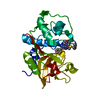
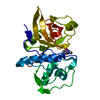
SHEET THE SHEET STRUCTURE OF THIS MOLECULE IS BIFURCATED. IN ORDER TO REPRESENT THIS FEATURE IN ... SHEET THE SHEET STRUCTURE OF THIS MOLECULE IS BIFURCATED. IN ORDER TO REPRESENT THIS FEATURE IN THE SHEET RECORDS BELOW, TWO SHEETS ARE DEFINED.

-
Structure visualization

Structure viewerMolecule:
MolmilJmol/JSmol

Downloads & links

-
Assembly

Deposited unit
A: CATHEPSIN S


Theoretical massNumber of molelcules
Total (without water)23,9761
Polymers23,9761
Non-polymers00
Water3,117173
1


  • Idetical with deposited unit
  • defined by author&software
TypeNameSymmetry operationNumber
identity operation1_555x,y,z1
MethodPQS
Unit cell
Length a, b, c (Å)79.989, 79.989, 61.517
Angle α, β, γ (deg.)90.00, 90.00, 120.00
Int Tables number152
Space group name H-MP3121

-
Components

#1: Protein CATHEPSIN S


Mass: 23975.900 Da / Num. of mol.: 1 / Mutation: YES
Source method: isolated from a genetically manipulated source
Source: (gene. exp.) HOMO SAPIENS (human) / Cell line (production host): SF9 / Production host: SPODOPTERA FRUGIPERDA (fall armyworm) / References: UniProt: P25774, cathepsin S
#2: Water ChemComp-HOH / water


Mass: 18.015 Da / Num. of mol.: 173 / Source method: isolated from a natural source / Formula: H2O
Compound detailsCHAIN A ENGINEERED MUTATION CYS139SER
Sequence detailsREFERENCE: WIEDERANDERS B, ET AL., (1992) J. BIOL. CHEM. 267 PP 13708-13713. THE SEQUENCE USED IS ...REFERENCE: WIEDERANDERS B, ET AL., (1992) J. BIOL. CHEM. 267 PP 13708-13713. THE SEQUENCE USED IS THAT PUBLISHED IN THE ABOVE REFERENCE THIS ACCOUNTS FOR THE TWO CONFLICTS NOTED IN THE SEQADV RECORDS

-
Experimental details

-
Experiment

ExperimentMethod: X-RAY DIFFRACTION / Number of used crystals: 1

-
Sample preparation

CrystalDensity Matthews: 2.3 Å3/Da / Density % sol: 46 %
Crystal growMethod: vapor diffusion, hanging drop / pH: 4.1
Details: CRYSTALLISATION WAS PERFORMED BY THE HANGING-DROP VAPOUR DIFFUSION METHOD. EQUAL VOLUMES CATHEPSIN S (CYS25SER) AT 7 MG/ML, INCLUDING THE PEPTIDE ABZ-LEU-THR-BAL-HYP-TYR(NO2)-ASP-NH2 AT 1 MM ...Details: CRYSTALLISATION WAS PERFORMED BY THE HANGING-DROP VAPOUR DIFFUSION METHOD. EQUAL VOLUMES CATHEPSIN S (CYS25SER) AT 7 MG/ML, INCLUDING THE PEPTIDE ABZ-LEU-THR-BAL-HYP-TYR(NO2)-ASP-NH2 AT 1 MM AND WELL SOLUTION WERE COMBINED AND PLACED OVER A WELL CONTAINING 20% ISOPROPANOL, 20% PEG2000 AND 0.1M SODIUM CITRATE, PH 4.13
Crystal grow
*PLUS
pH: 4.13 / Method: vapor diffusion, hanging drop
Components of the solutions
*PLUS
IDConc.Common nameCrystal-IDSol-IDDetails
17 mg/mlcathepsinS1drop
21 mMpeptide1drop
320 %2-propanol1reservoir
420 %PEG20001reservoir
50.1 Msodium citrate1reservoirpH4.13

-
Data collection

DiffractionMean temperature: 120 K
Diffraction sourceSource: ROTATING ANODE / Type: RIGAKU RUH3R / Wavelength: 1.5418
DetectorType: MARRESEARCH / Detector: IMAGE PLATE / Details: MULTILAYER
RadiationProtocol: SINGLE WAVELENGTH / Monochromatic (M) / Laue (L): M / Scattering type: x-ray
Radiation wavelengthWavelength: 1.5418 Å / Relative weight: 1
ReflectionResolution: 2.2→20 Å / Num. obs: 11969 / % possible obs: 99.4 % / Redundancy: 3.3 % / Rmerge(I) obs: 0.075 / Net I/σ(I): 16
Reflection shellResolution: 2.2→2.24 Å / Redundancy: 3 % / Rmerge(I) obs: 0.27 / Mean I/σ(I) obs: 4.5 / % possible all: 98.8
Reflection
*PLUS
Lowest resolution: 20 Å / Num. measured all: 39038
Reflection shell
*PLUS
Highest resolution: 2.2 Å / % possible obs: 98.8 % / Rmerge(I) obs: 0.27

-
Processing

Software
NameVersionClassification
REFMAC5refinement
DENZOdata reduction
SCALEPACKdata scaling
AMoREphasing
RefinementMethod to determine structure: MOLECULAR REPLACEMENT
Starting model: PDB ENTRY 1MEM
Resolution: 2.2→20 Å / Cor.coef. Fo:Fc: 0.933 / Cor.coef. Fo:Fc free: 0.887 / SU B: 6.157 / SU ML: 0.16 / Cross valid method: THROUGHOUT / ESU R: 0.3 / ESU R Free: 0.23 / Stereochemistry target values: MAXIMUM LIKELIHOOD
RfactorNum. reflection% reflectionSelection details
Rfree0.253 597 5.1 %RANDOM
Rwork0.193 ---
obs0.196 11182 99.5 %-
Solvent computationIon probe radii: 0.8 Å / Shrinkage radii: 0.8 Å / VDW probe radii: 1.4 Å / Solvent model: BABINET MODEL WITH MASK
Displacement parametersBiso mean: 19.04 Å2
Baniso -1Baniso -2Baniso -3
1-0.04 Å20.02 Å20 Å2
2--0.04 Å20 Å2
3----0.06 Å2
Refinement stepCycle: LAST / Resolution: 2.2→20 Å
ProteinNucleic acidLigandSolventTotal
Num. atoms1684 0 0 173 1857
Refine LS restraints
Refine-IDTypeDev idealDev ideal targetNumber
X-RAY DIFFRACTIONr_bond_refined_d0.0130.0211711
X-RAY DIFFRACTIONr_bond_other_d
X-RAY DIFFRACTIONr_angle_refined_deg
X-RAY DIFFRACTIONr_angle_other_deg1.6491.9422320
X-RAY DIFFRACTIONr_dihedral_angle_1_deg
X-RAY DIFFRACTIONr_dihedral_angle_2_deg
X-RAY DIFFRACTIONr_dihedral_angle_3_deg
X-RAY DIFFRACTIONr_dihedral_angle_4_deg
X-RAY DIFFRACTIONr_chiral_restr0.1150.2239
X-RAY DIFFRACTIONr_gen_planes_refined0.0060.021328
X-RAY DIFFRACTIONr_gen_planes_other
X-RAY DIFFRACTIONr_nbd_refined0.2340.3890
X-RAY DIFFRACTIONr_nbd_other
X-RAY DIFFRACTIONr_nbtor_refined
X-RAY DIFFRACTIONr_nbtor_other
X-RAY DIFFRACTIONr_xyhbond_nbd_refined0.1540.5193
X-RAY DIFFRACTIONr_xyhbond_nbd_other
X-RAY DIFFRACTIONr_metal_ion_refined
X-RAY DIFFRACTIONr_metal_ion_other
X-RAY DIFFRACTIONr_symmetry_vdw_refined0.2340.338
X-RAY DIFFRACTIONr_symmetry_vdw_other
X-RAY DIFFRACTIONr_symmetry_hbond_refined0.360.512
X-RAY DIFFRACTIONr_symmetry_hbond_other
X-RAY DIFFRACTIONr_symmetry_metal_ion_refined
X-RAY DIFFRACTIONr_symmetry_metal_ion_other
X-RAY DIFFRACTIONr_mcbond_it0.7391.51069
X-RAY DIFFRACTIONr_mcbond_other
X-RAY DIFFRACTIONr_mcangle_it1.37821700
X-RAY DIFFRACTIONr_mcangle_other
X-RAY DIFFRACTIONr_scbond_it2.2893642
X-RAY DIFFRACTIONr_scbond_other
X-RAY DIFFRACTIONr_scangle_it3.7124.5620
X-RAY DIFFRACTIONr_scangle_other
X-RAY DIFFRACTIONr_long_range_B_refined
X-RAY DIFFRACTIONr_long_range_B_other
X-RAY DIFFRACTIONr_rigid_bond_restr
X-RAY DIFFRACTIONr_sphericity_free
X-RAY DIFFRACTIONr_sphericity_bonded
LS refinement shellResolution: 2.2→2.26 Å / Total num. of bins used: 20 /
RfactorNum. reflection
Rfree0.336 41
Rwork0.206 790
Refinement
*PLUS
% reflection Rfree: 5 % / Rfactor obs: 0.197 / Rfactor Rfree: 0.251 / Rfactor Rwork: 0.194
Solvent computation
*PLUS
Displacement parameters
*PLUS
Refine LS restraints
*PLUS
Refine-IDTypeDev idealDev ideal target
X-RAY DIFFRACTIONr_bond_d0.0130.021
X-RAY DIFFRACTIONr_angle_d
X-RAY DIFFRACTIONr_angle_deg1.7541.942
X-RAY DIFFRACTIONr_plane_restr0.0060.02
X-RAY DIFFRACTIONr_chiral_restr0.1210.2

+
About Yorodumi

-
News

-
Feb 9, 2022. New format data for meta-information of EMDB entries

New format data for meta-information of EMDB entries

  • Version 3 of the EMDB header file is now the official format.
  • The previous official version 1.9 will be removed from the archive.

Related info.:EMDB header

External links:wwPDB to switch to version 3 of the EMDB data model

-
Aug 12, 2020. Covid-19 info

Covid-19 info

URL: https://pdbjlvh1.pdbj.org/emnavi/covid19.php

New page: Covid-19 featured information page in EM Navigator.

Related info.:Covid-19 info / Mar 5, 2020. Novel coronavirus structure data

+
Mar 5, 2020. Novel coronavirus structure data

Novel coronavirus structure data

Related info.:Yorodumi Speices / Aug 12, 2020. Covid-19 info

External links:COVID-19 featured content - PDBj / Molecule of the Month (242):Coronavirus Proteases

+
Jan 31, 2019. EMDB accession codes are about to change! (news from PDBe EMDB page)

EMDB accession codes are about to change! (news from PDBe EMDB page)

  • The allocation of 4 digits for EMDB accession codes will soon come to an end. Whilst these codes will remain in use, new EMDB accession codes will include an additional digit and will expand incrementally as the available range of codes is exhausted. The current 4-digit format prefixed with “EMD-” (i.e. EMD-XXXX) will advance to a 5-digit format (i.e. EMD-XXXXX), and so on. It is currently estimated that the 4-digit codes will be depleted around Spring 2019, at which point the 5-digit format will come into force.
  • The EM Navigator/Yorodumi systems omit the EMD- prefix.

Related info.:Q: What is EMD? / ID/Accession-code notation in Yorodumi/EM Navigator

External links:EMDB Accession Codes are Changing Soon! / Contact to PDBj

+
Jul 12, 2017. Major update of PDB

Major update of PDB

  • wwPDB released updated PDB data conforming to the new PDBx/mmCIF dictionary.
  • This is a major update changing the version number from 4 to 5, and with Remediation, in which all the entries are updated.
  • In this update, many items about electron microscopy experimental information are reorganized (e.g. em_software).
  • Now, EM Navigator and Yorodumi are based on the updated data.

External links:wwPDB Remediation / Enriched Model Files Conforming to OneDep Data Standards Now Available in the PDB FTP Archive

-
Yorodumi

Thousand views of thousand structures

  • Yorodumi is a browser for structure data from EMDB, PDB, SASBDB, etc.
  • This page is also the successor to EM Navigator detail page, and also detail information page/front-end page for Omokage search.
  • The word "yorodu" (or yorozu) is an old Japanese word meaning "ten thousand". "mi" (miru) is to see.

Related info.:EMDB / PDB / SASBDB / Comparison of 3 databanks / Yorodumi Search / Aug 31, 2016. New EM Navigator & Yorodumi / Yorodumi Papers / Jmol/JSmol / Function and homology information / Changes in new EM Navigator and Yorodumi

Read more