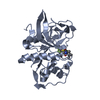
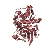
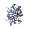
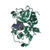
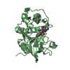
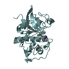
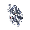
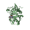
- PDB-4bs6: MOUSE CATHEPSIN S WITH COVALENT LIGAND -
+
Open data
ID or keywords:
Loading...
-
Basic information
Entry
Database: PDB / ID: 4bs6
Title
MOUSE CATHEPSIN S WITH COVALENT LIGAND
Components
CATHEPSIN S
Keywords
HYDROLASE / CYSTEINE PROTEASE / COVALENT LIGAND
Function / homology
Function and homology information
Assembly of collagen fibrils and other multimeric structures / Trafficking and processing of endosomal TLR / cathepsin S / regulation of antigen processing and presentation / Degradation of the extracellular matrix / basement membrane disassembly / Degradation of CDH1 / sensory perception of itch / antigen processing and presentation of peptide antigen / positive regulation of cation channel activity ...Assembly of collagen fibrils and other multimeric structures / Trafficking and processing of endosomal TLR / cathepsin S / regulation of antigen processing and presentation / Degradation of the extracellular matrix / basement membrane disassembly / Degradation of CDH1 / sensory perception of itch / antigen processing and presentation of peptide antigen / positive regulation of cation channel activity / MHC class II antigen presentation / proteoglycan binding / response to acidic pH / collagen catabolic process / fibronectin binding / bone resorption / collagen binding / phagocytic vesicle / Neutrophil degranulation / cysteine-type peptidase activity / laminin binding / early endosome lumen / : / protein processing / antigen processing and presentation of exogenous peptide antigen via MHC class II / positive regulation of inflammatory response / late endosome / peptidase activity / lysosome / cysteine-type endopeptidase activity / cell surface / proteolysis / : / membrane Similarity search - Function
Cathepsin propeptide inhibitor domain (I29) / Cathepsin propeptide inhibitor domain (I29) / Cathepsin propeptide inhibitor domain (I29) / Papain-like cysteine endopeptidase / Cysteine peptidase, asparagine active site / Eukaryotic thiol (cysteine) proteases asparagine active site. / Cysteine peptidase, histidine active site / Eukaryotic thiol (cysteine) proteases histidine active site. / : / Peptidase C1A, papain C-terminal ...Cathepsin propeptide inhibitor domain (I29) / Cathepsin propeptide inhibitor domain (I29) / Cathepsin propeptide inhibitor domain (I29) / Papain-like cysteine endopeptidase / Cysteine peptidase, asparagine active site / Eukaryotic thiol (cysteine) proteases asparagine active site. / Cysteine peptidase, histidine active site / Eukaryotic thiol (cysteine) proteases histidine active site. / : / Peptidase C1A, papain C-terminal / Papain family cysteine protease / Papain family cysteine protease / Cysteine proteinases / Cysteine peptidase, cysteine active site / Eukaryotic thiol (cysteine) proteases cysteine active site. / Cathepsin B; Chain A / Papain-like cysteine peptidase superfamily / Alpha-Beta Complex / Alpha Beta Similarity search - Domain/homology
Protocol: SINGLE WAVELENGTH / Monochromatic (M) / Laue (L): M / Scattering type: x-ray
Radiation wavelength
Wavelength: 1 Å / Relative weight: 1
Reflection
Resolution: 1.1→19.1 Å / Num. obs: 143497 / % possible obs: 82.9 % / Observed criterion σ(I): -3 / Redundancy: 2.74 % / Rmerge(I) obs: 0.04 / Net I/σ(I): 14.07
Reflection shell
Resolution: 1.1→1.2 Å / Redundancy: 0.73 % / Rmerge(I) obs: 0.42 / Mean I/σ(I) obs: 1.53 / % possible all: 36.8
-
Processing
Software
Name
Version
Classification
REFMAC
5.7.0029
refinement
XDS
datareduction
SADABS
datascaling
PHASER
phasing
Refinement
Method to determine structure: MOLECULAR REPLACEMENT Starting model: IN HOUSE STRUCTURES Resolution: 1.2→19.15 Å / Cor.coef. Fo:Fc: 0.983 / Cor.coef. Fo:Fc free: 0.975 / SU B: 1.129 / SU ML: 0.022 / Cross valid method: THROUGHOUT / ESU R: 0.034 / ESU R Free: 0.036 / Stereochemistry target values: MAXIMUM LIKELIHOOD / Details: HYDROGENS HAVE BEEN ADDED IN THE RIDING POSITIONS.
Rfactor
Num. reflection
% reflection
Selection details
Rfree
0.15497
6353
5.1 %
RANDOM
Rwork
0.12193
-
-
-
obs
0.12359
119007
93.8 %
-
Solvent computation
Ion probe radii: 0.8 Å / Shrinkage radii: 0.8 Å / VDW probe radii: 1.2 Å / Solvent model: MASK
 Movie
Movie Controller


 Open data
 Basic information
 Components
 Keywords
 Function and homology information
 X-RAY DIFFRACTION /
  Authors
 Citation
 Structure visualization
 Downloads & links
 Other downloads
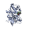


 PDBj
PDBj


 Assembly
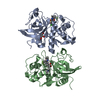


 Mass: 92.094 Da / Num. of mol.: 1 / Source method: obtained synthetically / Formula: C3H8O3
 Mass: 604.085 Da / Num. of mol.: 2 / Source method: obtained synthetically / Formula: C26H33ClF3N5O4S
 Mass: 18.015 Da / Num. of mol.: 702 / Source method: isolated from a natural source / Formula: H2O
 Sample preparation
 / Beamline: X10SA / Wavelength: 1
  Processing