 Movie
Movie Controller

[English] 日本語
 Yorodumi
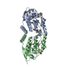
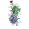
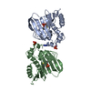
- PDB-5t6u: Crystal structure of mouse cathepsin K at 2.9 Angstroms resolution. -
+ Open data

- Basic information
| Entry | Database: PDB / ID: 5t6u | |||||||||
|---|---|---|---|---|---|---|---|---|---|---|
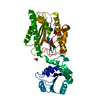
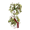
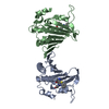
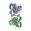
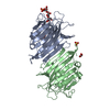
| Title | Crystal structure of mouse cathepsin K at 2.9 Angstroms resolution. | |||||||||
 Components Components | Cathepsin K | |||||||||
 Keywords Keywords | HYDROLASE / cathepsin K / N-ACETYL-D-GLUCOSAMINE | |||||||||
| Function / homology |  Function and homology information Function and homology informationRUNX1 regulates transcription of genes involved in differentiation of keratinocytes / Activation of Matrix Metalloproteinases / Collagen degradation / Trafficking and processing of endosomal TLR / cathepsin K / Degradation of the extracellular matrix / negative regulation of cartilage development / MHC class II antigen presentation / thyroid hormone generation / proteoglycan binding ...RUNX1 regulates transcription of genes involved in differentiation of keratinocytes / Activation of Matrix Metalloproteinases / Collagen degradation / Trafficking and processing of endosomal TLR / cathepsin K / Degradation of the extracellular matrix / negative regulation of cartilage development / MHC class II antigen presentation / thyroid hormone generation / proteoglycan binding / collagen catabolic process / fibronectin binding / bone resorption / collagen binding / cysteine-type peptidase activity / : / peptidase activity / lysosome / apical plasma membrane / external side of plasma membrane / cysteine-type endopeptidase activity / proteolysis / : / nucleoplasm Similarity search - Function | |||||||||
| Biological species |  | |||||||||
| Method |  X-RAY DIFFRACTION / SYNCHROTRON / MOLECULAR REPLACEMENT / Resolution: 2.9 Å X-RAY DIFFRACTION / SYNCHROTRON / MOLECULAR REPLACEMENT / Resolution: 2.9 Å | |||||||||
 Authors Authors | Law, S. / Aguda, A. / Nguyen, N. / Brayer, G. / Bromme, D. | |||||||||
| Funding support |  Canada, 2items Canada, 2items
| |||||||||
 Citation Citation | Journal: Biochem. J. / Year: 2017 Title: Identification of mouse cathepsin K structural elements that regulate the potency of odanacatib. Authors: Law, S. / Andrault, P.M. / Aguda, A.H. / Nguyen, N.T. / Kruglyak, N. / Brayer, G.D. / Bromme, D. | |||||||||
| History |
|
- Structure visualization
| Structure viewer | Molecule: MolmilJmol/JSmol |
|---|
- Downloads & links
-Download
| PDBx/mmCIF format | 5t6u.cif.gz | 55.7 KB | Display | PDBx/mmCIF format |
|---|---|---|---|---|
| PDB format | pdb5t6u.ent.gz | 38.4 KB | Display | PDB format |
| PDBx/mmJSON format | 5t6u.json.gz | Tree view | PDBx/mmJSON format | |
| Others |  Other downloads Other downloads |
-Validation report
| Arichive directory | https://data.pdbj.org/pub/pdb/validation_reports/t6/5t6uftp://data.pdbj.org/pub/pdb/validation_reports/t6/5t6u | HTTPS FTP |
|---|
-Related structure data
| Related structure data |  5tdiC  5tunC 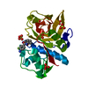 4x6hS S: Starting model for refinement C: citing same article ( |
|---|---|
| Similar structure data |


-Links
 PDBj
PDBj


- Assembly
| Deposited unit | 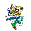
| ||||||||
|---|---|---|---|---|---|---|---|---|---|
| 1 | 
| ||||||||
| Unit cell |
| ||||||||
| Components on special symmetry positions |
|
-Components
| #1: Protein | Mass: 23448.391 Da / Num. of mol.: 1 Source method: isolated from a genetically manipulated source Source: (gene. exp.)  Komagataella pastoris GS115 (fungus) / References: UniProt: P55097, cathepsin K Komagataella pastoris GS115 (fungus) / References: UniProt: P55097, cathepsin K | ||||||
|---|---|---|---|---|---|---|---|
| #2: Chemical |   Mass: 96.063 Da / Num. of mol.: 3 / Source method: obtained synthetically / Formula: SO4 Mass: 96.063 Da / Num. of mol.: 3 / Source method: obtained synthetically / Formula: SO4#3: Sugar | ChemComp-NAG / |   Type: D-saccharide, beta linking / Mass: 221.208 Da / Num. of mol.: 1 Type: D-saccharide, beta linking / Mass: 221.208 Da / Num. of mol.: 1Source method: isolated from a genetically manipulated source Formula: C8H15NO6 #4: Water | ChemComp-HOH / |  Mass: 18.015 Da / Num. of mol.: 22 / Source method: isolated from a natural source / Formula: H2O Mass: 18.015 Da / Num. of mol.: 22 / Source method: isolated from a natural source / Formula: H2OHas protein modification | Y | |
-Experimental details
-Experiment
| Experiment | Method: X-RAY DIFFRACTION / Number of used crystals: 1 |
|---|
- Sample preparation
| Crystal | Density Matthews: 2.34 Å3/Da / Density % sol: 47.51 % |
|---|---|
| Crystal grow | Temperature: 298 K / Method: vapor diffusion, sitting drop / pH: 6.2 Details: 0.1 M sodium phosphate pH 6.2 25% (v/v) 1,2-propanediol 10% (v/v) glycerol |
-Data collection
| Diffraction | Mean temperature: 100 K |
|---|---|
| Diffraction source | Source: SYNCHROTRON / Site: SSRL  / Beamline: BL12-2 / Wavelength: 0.98 Å / Beamline: BL12-2 / Wavelength: 0.98 Å |
| Detector | Type: DECTRIS PILATUS 6M / Detector: PIXEL / Date: May 14, 2016 |
| Radiation | Protocol: SINGLE WAVELENGTH / Monochromatic (M) / Laue (L): M / Scattering type: x-ray |
| Radiation wavelength | Wavelength: 0.98 Å / Relative weight: 1 |
| Reflection | Resolution: 2.9→43.26 Å / Num. obs: 5243 / % possible obs: 99.53 % / Redundancy: 5.4 % / CC1/2: 0.99 / Rmerge(I) obs: 0.122 / Net I/av σ(I): 10.6 / Net I/σ(I): 10.66 |
| Reflection shell | Resolution: 2.9→3.06 Å / Redundancy: 5.8 % / Rmerge(I) obs: 0.32 / Mean I/σ(I) obs: 5.3 / CC1/2: 0.953 / % possible all: 99.9 |
- Processing
| Software |
| ||||||||||||||||||||||||
|---|---|---|---|---|---|---|---|---|---|---|---|---|---|---|---|---|---|---|---|---|---|---|---|---|---|
| Refinement | Method to determine structure: MOLECULAR REPLACEMENT Starting model: 4X6H Resolution: 2.9→43.26 Å / SU ML: 0.14 / Cross valid method: FREE R-VALUE / σ(F): 1.35 / Phase error: 19.45
| ||||||||||||||||||||||||
| Solvent computation | Shrinkage radii: 0.9 Å / VDW probe radii: 1.11 Å | ||||||||||||||||||||||||
| Refinement step | Cycle: LAST / Resolution: 2.9→43.26 Å
| ||||||||||||||||||||||||
| Refine LS restraints |
| ||||||||||||||||||||||||
| LS refinement shell |
|
