 Movie
Movie Controller

[English] 日本語
 Yorodumi
- PDB-1g7y: THE CRYSTAL STRUCTURE OF THE 58KD VEGETATIVE LECTIN FROM THE TROP... -
+ Open data

- Basic information
| Entry | Database: PDB / ID: 1g7y | |||||||||
|---|---|---|---|---|---|---|---|---|---|---|
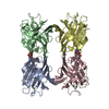
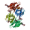
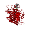
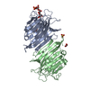
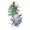
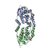
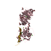
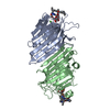
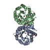
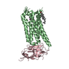
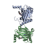
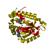
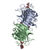
| Title | THE CRYSTAL STRUCTURE OF THE 58KD VEGETATIVE LECTIN FROM THE TROPICAL LEGUME DOLICHOS BIFLORUS | |||||||||
 Components Components | STEM/LEAF LECTIN DB58 | |||||||||
 Keywords Keywords | SUGAR BINDING PROTEIN / JELLY ROLL FOLD / LECTIN | |||||||||
| Function / homology |  Function and homology information Function and homology information | |||||||||
| Biological species |  Vigna unguiculata subsp. cylindrica (catjang cowpea) Vigna unguiculata subsp. cylindrica (catjang cowpea) | |||||||||
| Method |  X-RAY DIFFRACTION / SYNCHROTRON / MOLECULAR REPLACEMENT / Resolution: 2.5 Å X-RAY DIFFRACTION / SYNCHROTRON / MOLECULAR REPLACEMENT / Resolution: 2.5 Å | |||||||||
 Authors Authors | Buts, L. / Hamelryck, T.W. / Loris, R. / Dao-Thi, M.-H. / Wyns, L. / Etzler, M.E. | |||||||||
 Citation Citation | Journal: J.Mol.Biol. / Year: 2001 Title: Weak protein-protein interactions in lectins: the crystal structure of a vegetative lectin from the legume Dolichos biflorus. Authors: Buts, L. / Dao-Thi, M.H. / Loris, R. / Wyns, L. / Etzler, M. / Hamelryck, T. #1: Journal: Acta Crystallogr.,Sect.D / Year: 1998Title: Crystallisation of two related lectins from the legume plant Dolichos biflorus Authors: Dao-Thi, M. / Hamelryck, T.W. / Bouckaert, J. / Koerber, F. / Burkow, V. / Poortmans, F. / Etzler, M. / Strecker, G. / Wyns, L. / Loris, R. #2: Journal: J.Mol.Biol. / Year: 1999Title: Carbohydrate binding, quaternary structure and a novel hydrophobic binding site in two legume lectins oligomers from Dolichos biflorus Authors: Hamelryck, T.W. / Loris, R. / Bouckaert, J. / Dao-Thi, M.-H. / Strecker, G. / Imberty, A. / Fernandez, E. / Wyns, L. / Etzler, M.E. | |||||||||
| History |
|
- Structure visualization
| Structure viewer | Molecule: MolmilJmol/JSmol |
|---|
- Downloads & links
-Download
| PDBx/mmCIF format | 1g7y.cif.gz | 287.4 KB | Display | PDBx/mmCIF format |
|---|---|---|---|---|
| PDB format | pdb1g7y.ent.gz | 230.5 KB | Display | PDB format |
| PDBx/mmJSON format | 1g7y.json.gz | Tree view | PDBx/mmJSON format | |
| Others |  Other downloads Other downloads |
-Validation report
| Arichive directory | https://data.pdbj.org/pub/pdb/validation_reports/g7/1g7yftp://data.pdbj.org/pub/pdb/validation_reports/g7/1g7y | HTTPS FTP |
|---|
-Related structure data
-Links
 PDBj
PDBj

- Assembly
| Deposited unit | 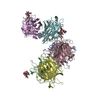
| ||||||||
|---|---|---|---|---|---|---|---|---|---|
| 1 | 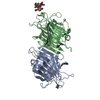
| ||||||||
| 2 | 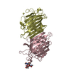
| ||||||||
| 3 | 
| ||||||||
| Unit cell |
|
-Components
-Protein , 1 types, 6 molecules ABCDEF
| #1: Protein | Mass: 27146.885 Da / Num. of mol.: 6 / Source method: isolated from a natural source / Details: TROPICAL LEGUME Source: (natural) Vigna unguiculata subsp. cylindrica (catjang cowpea)Species: Vigna unguiculata / Strain: subsp. cylindrica / References: UniProt: P19588 |
|---|
-Sugars , 2 types, 3 molecules
| #2: Polysaccharide | Source method: isolated from a genetically manipulated source #3: Polysaccharide | beta-L-fucopyranose-(1-3)-[2-acetamido-2-deoxy-beta-D-glucopyranose-(1-4)]2-acetamido-2-deoxy-beta- ...beta-L-fucopyranose-(1-3)-[2-acetamido-2-deoxy-beta-D-glucopyranose-(1-4)]2-acetamido-2-deoxy-beta-D-glucopyranose | Source method: isolated from a genetically manipulated source |
|---|
-Non-polymers , 3 types, 53 molecules 


| #4: Chemical | ChemComp-CA /  Mass: 40.078 Da / Num. of mol.: 6 / Source method: obtained synthetically / Formula: Ca Mass: 40.078 Da / Num. of mol.: 6 / Source method: obtained synthetically / Formula: Ca#5: Chemical | ChemComp-MN /  Mass: 54.938 Da / Num. of mol.: 6 / Source method: obtained synthetically / Formula: Mn Mass: 54.938 Da / Num. of mol.: 6 / Source method: obtained synthetically / Formula: Mn#6: Water | ChemComp-HOH / | Mass: 18.015 Da / Num. of mol.: 41 / Source method: isolated from a natural source / Formula: H2O |
|---|
-Details
| Has protein modification | Y |
|---|
-Experimental details
-Experiment
| Experiment | Method: X-RAY DIFFRACTION / Number of used crystals: 1 |
|---|
- Sample preparation
| Crystal | Density Matthews: 2.77 Å3/Da / Density % sol: 55.54 % | ||||||||||||||||||||||||||||||
|---|---|---|---|---|---|---|---|---|---|---|---|---|---|---|---|---|---|---|---|---|---|---|---|---|---|---|---|---|---|---|---|
| Crystal grow | Temperature: 293 K / Method: vapor diffusion, hanging drop / pH: 5.6 Details: 5-7 mg/ml protein; 100mM sodium citrate; 8-12%(w/v) PEG(6000); 0.2-0.4M sodium chloride, pH 5.6, VAPOR DIFFUSION, HANGING DROP, temperature 293K | ||||||||||||||||||||||||||||||
| Crystal grow | *PLUS | ||||||||||||||||||||||||||||||
| Components of the solutions | *PLUS
|
-Data collection
| Diffraction | Mean temperature: 100 K |
|---|---|
| Diffraction source | Source: SYNCHROTRON / Site: EMBL/DESY, HAMBURG  / Beamline: BW7A / Wavelength: 0.98089 Å / Beamline: BW7A / Wavelength: 0.98089 Å |
| Detector | Type: MARRESEARCH / Detector: IMAGE PLATE / Date: Aug 20, 1998 |
| Radiation | Monochromator: MIRRORS / Protocol: SINGLE WAVELENGTH / Monochromatic (M) / Laue (L): M / Scattering type: x-ray |
| Radiation wavelength | Wavelength: 0.98089 Å / Relative weight: 1 |
| Reflection | Resolution: 2.5→20 Å / Num. all: 276382 / Num. obs: 259246 / % possible obs: 93.8 % / Observed criterion σ(I): 5 / Redundancy: 4.4 % / Biso Wilson estimate: 45.1 Å2 / Rmerge(I) obs: 0.104 / Net I/σ(I): 12.9 |
| Reflection shell | Resolution: 2.5→2.59 Å / Rmerge(I) obs: 0.656 / Mean I/σ(I) obs: 2.78 / % possible all: 95.7 |
| Reflection | *PLUS Num. all: 259246 / Num. obs: 59075 / Num. measured all: 276382 |
| Reflection shell | *PLUS % possible obs: 95.7 % |
- Processing
| Software |
| ||||||||||||||||||||||||||||||||||||||||
|---|---|---|---|---|---|---|---|---|---|---|---|---|---|---|---|---|---|---|---|---|---|---|---|---|---|---|---|---|---|---|---|---|---|---|---|---|---|---|---|---|---|
| Refinement | Method to determine structure: MOLECULAR REPLACEMENT / Resolution: 2.5→20 Å / Rfactor Rfree error: 0.003 / Data cutoff high absF: 2558709.98 / Data cutoff low absF: 0 / Isotropic thermal model: RESTRAINED / Cross valid method: THROUGHOUT / σ(F): 0 / σ(I): 0 / Stereochemistry target values: Engh & Huber
| ||||||||||||||||||||||||||||||||||||||||
| Solvent computation | Solvent model: FLAT MODEL / Bsol: 31.12 Å2 / ksol: 0.289 e/Å3 | ||||||||||||||||||||||||||||||||||||||||
| Displacement parameters | Biso mean: 38.8 Å2
| ||||||||||||||||||||||||||||||||||||||||
| Refine analyze |
| ||||||||||||||||||||||||||||||||||||||||
| Refinement step | Cycle: LAST / Resolution: 2.5→20 Å
| ||||||||||||||||||||||||||||||||||||||||
| Refine LS restraints |
| ||||||||||||||||||||||||||||||||||||||||
| Refine LS restraints NCS | NCS model details: CONSTR | ||||||||||||||||||||||||||||||||||||||||
| LS refinement shell | Resolution: 2.5→2.66 Å / Rfactor Rfree error: 0.011 / Total num. of bins used: 6
| ||||||||||||||||||||||||||||||||||||||||
| Xplor file |
| ||||||||||||||||||||||||||||||||||||||||
| Software | *PLUS Name: CNS / Version: 1 / Classification: refinement | ||||||||||||||||||||||||||||||||||||||||
| Refinement | *PLUS Lowest resolution: 20 Å / σ(F): 0 / % reflection Rfree: 10.1 % | ||||||||||||||||||||||||||||||||||||||||
| Solvent computation | *PLUS | ||||||||||||||||||||||||||||||||||||||||
| Displacement parameters | *PLUS Biso mean: 38.8 Å2 | ||||||||||||||||||||||||||||||||||||||||
| Refine LS restraints | *PLUS
| ||||||||||||||||||||||||||||||||||||||||
| LS refinement shell | *PLUS Rfactor Rfree: 0.359 / % reflection Rfree: 10 % / Rfactor Rwork: 0.314 |
